
Ever looked at a chest X-ray and thought, “Wait… am I supposed to see something here, or is this just modern art in black, white, and gray?” You’re not alone. A chest X-ray is one of the most common medical tests in the world, yet for most people, the results are downright baffling. Then, to make it even more confusing, the report might simply say: “Normal findings.” And you’re left wondering—what does a normal chest X-ray even look like? And how is it different from something serious like pneumonia?
That’s where this guide comes in. Think of it as your friendly decoder. We’ll walk through what a normal chest X-ray should show, what a typical report says (without the scary medical jargon), and the key differences between normal and abnormal images. By the end, you’ll be able to look at a chest X-ray with a bit more confidence instead of squinting at it like a puzzle you can’t solve.
Of course, all of this clarity starts with one crucial factor: quality. A crisp, high-quality image makes all the difference between “Hmm, maybe that’s the lung?” and an accurate, reliable diagnosis. That’s why medical professionals rely on advanced imaging films like HSIN Film’s medical dry film—because when it comes to your health, blurry just doesn’t cut it.
So, let’s dive in and finally make sense of the mysterious world of chest X-rays—minus the guesswork and confusion.
Table of Contents
What a “Normal View” Looks Like and Its Basic Findings
So, let’s clear up one of the first mysteries: what does a normal chest X-ray actually look like? If you’ve ever seen one hanging up in a doctor’s office, you’ve probably noticed that it’s not exactly intuitive—there’s no big neon sign pointing at the lungs saying, “Yep, these are fine.” But there is a method to the madness.
The Standard View: PA (Posterior-Anterior)
The most common view—and the gold standard—is called the Posterior-Anterior (PA) view. Here’s what that means in normal-people terms: the X-ray beam passes from your back through to your chest. This positioning isn’t random—it ensures the heart and lungs are displayed in the most accurate way possible. Think of it like taking a passport photo: the rules may feel picky, but the consistency means professionals can compare apples to apples.
Fun fact: according to radiology studies, the PA view reduces distortion of the heart size compared to the AP (Anterior-Posterior) view, which is why radiologists prefer it for evaluating normal chest X-rays.
Reading the Shades
Now, here’s where the puzzle comes in. A chest X-ray is basically a black-and-white snapshot of what’s inside your chest, and every shade tells a story:
- Black = Air. This is where your lungs and trachea show up. The darker the space, the more air there is, which is a good thing (unless it’s in the wrong place, like outside the lungs).
- Gray = Soft Tissues. Your heart, blood vessels, and other soft tissues appear in gray. This is the “middle ground” where radiologists pay close attention to shape and borders.
- White = Bones. Dense structures like ribs, clavicles, and the spine soak up more radiation, so they show up bright white. Basically, they’re the high-contrast outlines in the image.
Pro tip: Once you know this black-gray-white system, X-rays stop looking like abstract art and start making sense.
Normal Findings in a Healthy Chest
So, what does a radiologist expect to see in a normal chest X-ray? Here’s the checklist:
- Clear, well-aerated lungs. No foggy patches, no suspicious shadows—just nice, dark spaces showing healthy airflow.
- A heart of normal size and shape. Fun stat: your heart should be less than half the width of your chest on a PA view. Any bigger, and radiologists start thinking about conditions like cardiomegaly (an enlarged heart).
- Intact bones. The ribs, spine, and collarbones should look continuous and unbroken—no cracks, no odd bends.
Think of it like evaluating a car from the outside: the tires (bones) should be aligned, the windshield (lungs) should be clear, and the engine block (heart) should be the right size—not bulging out of the hood.
A Detailed Look at the Normal Chest X-ray
Okay, so now that we’ve got the basics down, let’s zoom in and take a detailed tour of what a normal chest X-ray actually shows. Imagine we’re walking through a map of your chest, one landmark at a time.
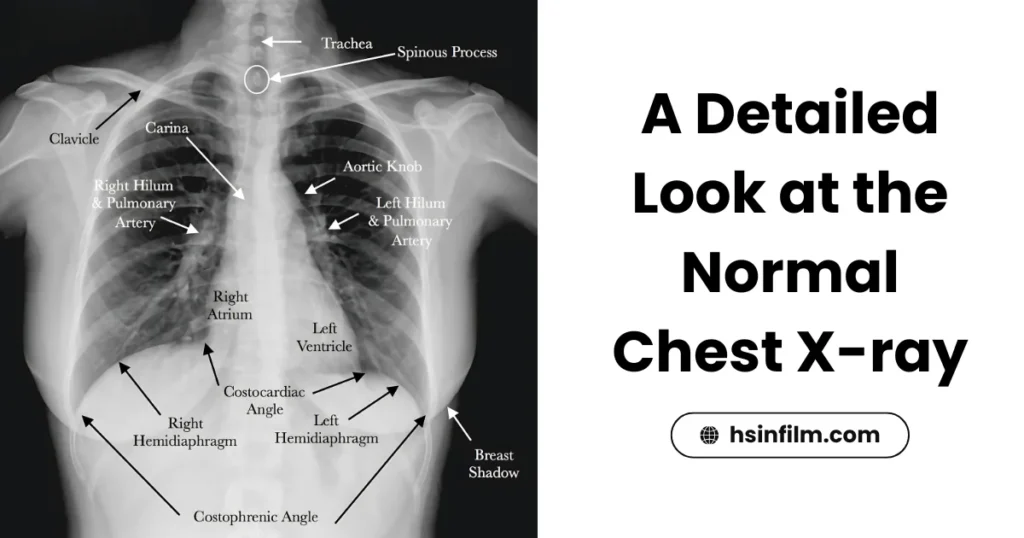
The Lungs: Big, Black, and Beautiful
First stop: the lungs. On a normal chest X-ray, the lungs show up as large, symmetrical black spaces—that’s the air doing its job. Don’t worry if you see thin white lines weaving through the black background; those are just your blood vessels and airways, totally normal. Think of it like city streets showing up on a satellite map—you want to see them, because they prove the neighborhood (your lungs) is alive and functioning.
If the lungs look cloudy, patchy, or uneven, that’s when radiologists start suspecting conditions like pneumonia or fluid buildup. But in a normal scan, it’s clear skies all the way.
The Heart: Center Stage, but Not Too Big
The heart shows up as a soft, whitish shadow right in the middle of the chest. Here’s the key: on a normal chest X-ray, the heart should take up less than half the width of the ribcage. Any larger, and doctors start thinking about issues like an enlarged heart.
For women, there’s an extra layer: breast tissue. On X-rays, this shows up as a hazy, soft shadow over the lower lung fields. It’s a completely normal finding but can sometimes trick the untrained eye into thinking there’s a problem. Radiologists are trained to spot this so they don’t confuse normal anatomy with disease.
The Bones: Bright and Bold
Next, check out the skeleton framework. The ribs, collarbones, and shoulder blades should appear sharp, clearly defined, and bright white. These are your structural supports. Radiologists scan them carefully for any cracks, breaks, or weird shapes. In a normal film, everything lines up perfectly, like the framing of a house.
The Diaphragm: The Smooth Domes
At the very bottom, you’ll spot the diaphragm—two smooth, curved white domes sitting beneath the lungs. On a normal chest X-ray, the right dome usually sits a little higher than the left (thanks to the liver pushing it up). Both should be well-defined and smooth, like calm waves at the edge of the ocean.
The Report: Turning Pictures Into Words
All of these findings—lungs that are clear, a heart that’s well-sized, bones that are intact, and a diaphragm that’s smooth—get translated into a written summary: your normal chest X-ray report. It’s the radiologist’s way of saying, “Everything looks good here, nothing unusual to worry about.”
So, the next time you get handed that short and slightly vague report, you’ll know exactly what’s hiding behind those words. Instead of staring at the image like it’s a medical Rorschach test, you’ll understand what “normal” really looks like—and more importantly, why that’s a good thing.
Normal vs. Abnormal: A Case Study with Pneumonia
Alright, now let’s get real. We’ve talked about what a normal chest X-ray looks like—clear lungs, neat bones, smooth diaphragm, and a heart that’s playing by the rules. But what happens when something’s not right? That’s where the word “abnormal” comes in.
Understanding Abnormal
An abnormal chest X-ray is basically a red flag. Instead of the clean, expected picture, radiologists might see:
- Fluid pooling in the lungs or around them.
- Growths or masses that shouldn’t be there.
- Broken bones interrupting the clean white lines of the ribs.
- An enlarged heart, taking up more space than it should.
In other words, anything that doesn’t match the blueprint of “normal” stands out like a coffee stain on a white shirt.
Normal vs. Pneumonia: The Classic Example
One of the most common and dramatic comparisons is normal chest X-ray vs. pneumonia. Here’s the difference in plain English:
- Normal Lung: Imagine a clear night sky—black, open, uncluttered. That’s what a healthy lung looks like on X-ray: big black spaces filled with air, maybe with some faint white lines (blood vessels) crisscrossing.
- Pneumonia: Now imagine clouds rolling across that sky. On X-ray, pneumonia shows up as white or patchy cloudy areas called “consolidation.” Why? Because the tiny air sacs in the lungs, which should be full of air, are now jam-packed with fluid, pus, and inflammation. Those dense contents block the X-ray beam, turning the area from black to white.
The Difference You Can See
The contrast between the jet-black air-filled lung in a normal chest X-ray and the cloudy, patchy white of pneumonia is huge—almost impossible to miss once you know what you’re looking for. Radiologists rely on that stark difference to confirm pneumonia and guide treatment.
In fact, research shows that chest X-rays remain one of the most reliable tools for diagnosing pneumonia, especially when combined with symptoms like fever, cough, and chest pain. It’s the medical equivalent of catching someone red-handed—the X-ray practically shouts, “Yep, there’s something wrong here.”
Real-Life Scenario
Picture this: a patient comes into the clinic with a high fever, bad cough, and shortness of breath. The doctor orders an X-ray. On the left side of the screen is the normal chest X-ray—clear and black. On the right? The same patient’s film, but this time, cloudy white patches cover part of the lung. That one image makes the difference between, “You’re fine, go home and rest” and “You’ve got pneumonia, let’s treat this right away.”
That’s the power of understanding the difference between normal and abnormal—it’s not just about shades of gray, it’s about catching disease before it gets worse.
Normal vs. Pneumonia on Chest X-ray
| Feature | Normal Chest X-ray | Chest X-ray with Pneumonia |
|---|---|---|
| Lung Appearance | Clear, black spaces filled with air | White or cloudy patches (consolidation) |
| Blood Vessels | Thin white lines visible, normal branching | Often obscured by dense white areas |
| Air Sacs (Alveoli) | Filled with air, invisible on X-ray | Filled with fluid/pus, block X-rays |
| Overall Pattern | Symmetrical, clean, well-aerated | Patchy, irregular, cloudy areas |
| Clinical Meaning | Healthy lungs, no infection | Active infection (pneumonia) requiring treatment |
The Normal Chest X-ray Report Format
Alright, so you’ve had your X-ray taken, you’ve stared at the mysterious black-and-white image, and now you’re handed… a one-page report. If you’ve ever looked at one and thought, “Wait, that’s it? This tiny paragraph tells me if I’m okay or not?” — you’re not alone. But here’s the thing: that report is the official record of the radiologist’s interpretation. It’s short, yes, but it’s also packed with meaning.
Purpose: Why the Report Matters
Think of the report as the translator between doctor-speak and decision-making. The image itself is just the raw data. The radiologist’s job is to analyze every detail and summarize it in clear, structured wording. For a normal chest X-ray, the report reassures both you and your doctor that everything looks healthy and no hidden surprises are lurking.
The Standard Format of a Normal Report
While reports can vary a bit from hospital to hospital, most follow the same core structure:
- Clinical Information
This is the who, why, and when of the exam. It includes patient details (name, age, medical ID) and the reason the X-ray was ordered (e.g., “Cough and fever for 5 days” or “Routine pre-employment screening”). - Findings
This is the meat of the report, where the radiologist goes system by system. For a normal chest X-ray, it often looks like a neat checklist:- Lungs are clear and well-aerated.
- Heart size is within normal limits.
- Bony thorax (ribs, spine, clavicles) is intact.
- Diaphragm is smooth and normally positioned.
- No abnormal fluid collections.
- Impression/Conclusion
This is the headline, the bolded summary, the “bottom line” moment. For a normal report, it usually reads something like: “No evidence of acute cardiopulmonary disease.” Translation? Your chest looks perfectly healthy. Nothing alarming, no urgent conditions—just business as usual.
Real-Life Example
Let’s say you went in for an X-ray because of a nagging cough. You get the report back, and under Impression it says: “Normal chest X-ray. No active pulmonary disease.” That single line tells your doctor: lungs are clear, no pneumonia, no fluid, no masses. In other words, it’s good news—you just might need some cough syrup, not antibiotics or hospital admission.
Conclusion
By now, you’ve taken a full tour of the normal chest X-ray—from the standard PA view and the shades of black, gray, and white, to the fine details of lungs, heart, bones, and diaphragm. You’ve also seen how a normal report is structured and how it contrasts with abnormal findings like pneumonia. This knowledge doesn’t just satisfy curiosity—it empowers you as a patient. When you understand what’s “normal,” you can better appreciate when something isn’t, and why your doctor recommends certain treatments.
At the heart of it all (pun intended), accurate diagnostics depend on one simple factor: image quality. A crystal-clear X-ray gives doctors the confidence to make the right call the first time, while a poor-quality film can create confusion, misdiagnosis, or unnecessary repeat scans. That’s why providers who use advanced solutions like HSIN’s high-quality dry films and printers are making an investment not just in technology—but in your health.
So the next time you see the words “Normal chest X-ray” on your report, you’ll know exactly what it means—and you can rest easy knowing that normal never looked so good.
For Clinics & Healthcare Providers: Accuracy starts with clarity. Upgrade your diagnostic imaging with HSIN’s medical dry films, thermal printers, and inkjet printers—engineered for sharp detail, durability, and consistent quality. With HSIN, every normal chest X-ray (and every abnormal one) tells the full story, the first time. Because when patients trust the image, they trust you.