Choosing the right film type shapes everything downstream — image quality, workflow speed, running costs, and how well your imaging equipment holds up over time. Yet many buyers default to whatever film their facility has always used, without ever comparing it against the alternatives. The differences between dry, thermal, and laser film are bigger than most people expect, and picking the wrong one for a given procedure can mean blurrier diagnostics, slower turnaround, or money spent on capability you don’t actually need.
This guide breaks down how each film type works, where each one fits best, and how to make a confident choice for your specific practice.
Table of Contents: Types of Medical X-ray Films
Why Film Type Still Matters
Digital imaging has reshaped much of modern radiology, but physical film hasn’t disappeared — it remains central to archiving, patient handoffs between facilities, surgical reference copies, and settings where digital infrastructure isn’t fully built out. Veterinary practices, smaller clinics, and facilities in regions still transitioning to fully digital PACS systems all continue to rely on film as a working part of their diagnostic process.
Not all film serves that role equally well. Dry, thermal, and laser film differ in how they’re exposed, how they’re processed, what equipment they require, and what kind of image they ultimately produce. Using the wrong type for a given printer or procedure isn’t just an inconvenience — it can mean streaky or low-contrast images, wasted sheets, and delays that ripple into patient care. Understanding what separates these three film types is the first step toward avoiding that.
Dry, Thermal & Laser Film Compared
| Film Type | How It Works | Contrast & Resolution | Best For | Advantages | Tradeoffs |
|---|---|---|---|---|---|
| Medical Dry Film | Heat-sensitive coating, no chemicals or darkroom | High contrast, moderate resolution | General radiology, portable/low-volume setups | Lowest running cost, simplest workflow, no chemical waste | Slightly lower resolution ceiling than laser |
| Medical Thermal Film | Direct thermal printing, instant dry output | Moderate-to-high contrast | Ultrasound, MRI, dental, high-throughput departments | Fastest turnaround, low maintenance, works with most thermal dry imagers | Sensitive to heat during storage; not the top choice for the finest diagnostic detail |
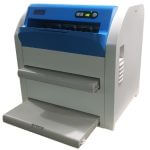
| Medical Laser Film | Laser-exposed silver halide emulsion | Highest contrast and resolution | CT, MRI, mammography, orthopedic imaging | Sharpest diagnostic clarity, most durable image | Highest cost per sheet; some laser imagers still require chemical development |
Medical Dry Film
Dry film is built around simplicity and cost control. It skips chemical developers, fixers, and darkroom processing entirely — the image forms through a heat-sensitive coating that reacts directly to a thermal print head, with no liquid chemistry involved at any stage. That translates into lower ongoing supply costs, less equipment maintenance, and a cleaner overall workflow, since there’s no developer to mix, no fixer to dispose of, and no darkroom to keep light-sealed.
This makes dry film a strong fit for general radiology work and for practices that don’t need to push resolution to its absolute limit on every image — smaller clinics, veterinary practices, and mobile or portable imaging setups in particular. It’s also a sensible default for facilities trying to control per-image costs across high sheet volumes, since eliminating chemical processing removes one of the more expensive recurring line items in a film-based workflow.
The tradeoff is resolution. Dry film produces perfectly serviceable diagnostic images for routine work, but it doesn’t quite match laser film when the finest anatomical detail is the deciding factor in a diagnosis.
Medical Thermal Film
Thermal film is built for speed. Using direct thermal printing, it produces a finished image almost instantly, with nothing — no ink, no toner, no chemical bath — standing between image capture and a usable printout. For departments where patient throughput is the primary constraint, that speed advantage compounds quickly: faster image turnaround means faster reads, faster patient discharge, and less bottlenecking at the imaging station itself.
This makes thermal film especially well-suited to busy ultrasound, MRI, and dental imaging workflows, where volume is high and the diagnostic requirement doesn’t call for laser-level resolution. It also tends to be compatible with a broad range of thermal dry imagers already in circulation, which can make it a straightforward drop-in replacement when switching suppliers.
The main limitation is sensitivity to heat and humidity, both during storage and in daily handling — more so than dry or laser film. A thermal sheet left in a warm storage room or handled with damp gloves is more likely to show quality loss than the other two film types. Proper storage (covered below) matters more here than anywhere else in this comparison.
Medical Laser Film
Laser film is the choice when diagnostic clarity can’t be compromised. A laser exposes a silver halide emulsion layer, producing the sharpest, highest-resolution image of the three film types under comparison here. That precision is why laser film remains the standard for CT, MRI, mammography, and orthopedic imaging — procedures where subtle density variations or fine structural detail can directly change a diagnosis or treatment plan.
That level of clarity comes at a cost, in more than one sense. Laser film is typically the most expensive per sheet of the three options, and depending on the specific imager in use, some laser systems still require a chemical development step — meaning the darkroom-processing considerations that dry and thermal film eliminate can still apply here.
For facilities where image resolution is the deciding factor — radiology departments handling complex diagnostic cases, or specialties like mammography where early detection depends on catching subtle abnormalities — the added cost of laser film is generally the right tradeoff.
Cost Considerations Beyond the Price Tag
Comparing film types purely on per-sheet price tells an incomplete story. A few other cost factors are worth weighing:
- Processing overhead: Dry and thermal film eliminate chemical costs entirely — no developer, fixer, or disposal fees. If your laser film system requires chemical processing, factor those recurring costs into the comparison, not just the film price itself.
- Reprint rate: A cheaper film that produces more unusable images due to lower resolution or handling sensitivity can end up costing more in wasted sheets and repeat imaging than a pricier, more forgiving option.
- Equipment maintenance: Chemical-based processing systems require more regular upkeep — cleaning, chemical replenishment, calibration — than dry thermal printers, which adds labor cost even when the film itself is inexpensive.
- Storage losses: Thermal film’s heat sensitivity means poor storage conditions can lead to a higher spoilage rate than dry or laser film, effectively raising its real-world cost if storage isn’t well controlled.
Looking at total cost per usable diagnostic image, rather than cost per sheet, usually gives a clearer picture of which film type is actually the most economical for a given practice.
Check our high quality and affordable medical X-ray films
Printer and Equipment Compatibility
Film type and printer compatibility are inseparable decisions — the wrong pairing simply won’t produce a usable image, regardless of film quality. Before switching or standardizing on a film type, confirm:
- Printer type: Dry thermal printers, direct thermal printers, and laser imagers each require film manufactured for that specific print method. Film and printer type are not interchangeable.
- DICOM and PACS compatibility: If your facility runs a DICOM-based digital workflow alongside film output, confirm the film and printer combination supports smooth digital-to-film integration.
- Sizing needs: Common sizes include 14×17, 11×14, 10×12, and 8×10 inches — make sure your chosen film is available in the sizes your imaging procedures actually require.
- Multi-brand compatibility: Many practices use film from a different manufacturer than their printer’s original brand as a cost-saving measure. Where that’s the plan, confirm compatibility explicitly rather than assuming — printer manufacturers don’t always publish full third-party compatibility lists.
How to Choose the Right Film for Your Practice
A handful of questions can narrow the decision quickly:
- Is turnaround speed your top priority? Thermal film’s near-instant output makes it the strongest fit for high-volume, fast-paced departments like ultrasound or busy dental practices.
- Is cost control the main driver? Dry film offers the lowest running cost, with no chemical processing required at any stage.
- Is maximum resolution essential? Laser film remains the standard where diagnostic precision can’t be compromised — CT, MRI, and mammography especially.
- Do you need more than one film type? Many practices don’t standardize on a single option — they mix film types by department, using laser film for CT/MRI, thermal film for high-throughput ultrasound, and dry film for general or portable radiology, matching each film to the procedure it serves best.
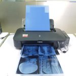
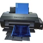
| HSIN Medical Dry Film | Best for inkjet printers, cost-saving |
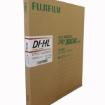
| HSIN Medical Thermal Film | Best for thermal dry imagers, fast turnaround |
| Medical Laser Film | Best for high-volume diagnostic clarity |
Handling and Processing Considerations
Even chemical-free film types benefit from careful handling practices:
- Dry and thermal film skip chemical processing altogether, which removes an entire category of quality risk — no developer temperature drift to manage, no fixer residue to worry about, no darkroom light leaks to guard against. The main handling risk instead shifts to the printer itself: keeping thermal printheads clean and well-maintained prevents streaking, banding, or uneven density in the final image.
- Laser film on chemical-based systems still carries the same sensitivities as traditional wet processing: development time and temperature directly affect image contrast, inadequate fixing can leave residual chemicals that cloud the image over time, and any light exposure in the darkroom before processing can fog the film and reduce diagnostic usability.
- Across all three film types, always handle unexposed and undeveloped film by the edges rather than the surface, avoid transferring oils or moisture from bare hands onto the imaging layer, and keep film shielded from ambient light until it’s actually processed.
Storing Your Film to Protect Image Quality
Proper storage extends the usable life of any film type and protects image quality well before the film is ever exposed:
- Temperature: Aim to store film between roughly 10–21°C (50–70°F). Excess heat can soften or degrade the coating — a particular risk for thermal film specifically — while excessively cold conditions can make film brittle and prone to physical damage.
- Humidity: Keep relative humidity in the 30–50% range. Too much humidity risks sticking, mold, or emulsion damage; too little can lead to static discharge that creates artifacts on the film before it’s even used.
- Light exposure: Store all film types in opaque, lightproof packaging until use — this is especially critical for laser film, given how light-sensitive its silver halide emulsion is.
- Handling and organization: Store film upright rather than flat or stacked, to avoid pressure damage over time. Use protective sleeves for anything not immediately in use, and keep a climate-controlled, organized archive for long-term storage rather than an ad hoc supply closet.
- Shelf life awareness: All film types have a finite shelf life even under ideal conditions. Rotate stock so older film gets used first, and avoid over-ordering beyond what your practice will use within its expected shelf-life window.
FAQ
Can I switch film types without changing my printer?
No — dry, thermal, and laser film each require a printer built for that specific print method. Switching film types generally means switching or adding compatible imaging equipment.
Which film type is cheapest to run long-term?
Dry film typically has the lowest total running cost, since it eliminates chemical processing costs and equipment maintenance associated with wet processing. Thermal film is close behind on cost but can incur higher losses from heat-sensitive storage issues if not handled carefully.
Do I have to use only one film type across my whole practice?
Not at all. Many practices assign film type by department or procedure — for example, laser film for CT and MRI, thermal film for high-volume ultrasound, and dry film for general radiography — rather than standardizing on a single option across the board.
Is laser film always the best choice for image quality?
It typically produces the sharpest, highest-resolution image of the three, which is why it’s the standard for CT, MRI, and mammography. But for routine radiography where that level of resolution isn’t required, dry or thermal film can be perfectly adequate and considerably more cost-effective.
Conclusion: Navigating the Dynamic World of X-Ray Films
Dry, thermal, and laser film each solve a different problem: dry film for cost-efficient simplicity, thermal film for speed and high-volume workflows, and laser film for maximum diagnostic clarity where the finest detail matters most. There’s no single “best” film type in the abstract — the right choice, or right combination of choices, comes down to matching film type to the specific procedure, equipment, and workflow priorities of your practice.