Orthodontic X-rays are a vital tool in dental care, providing detailed images of the teeth and jaw to aid in diagnosis and treatment planning. This comprehensive guide will help you understand the importance of orthodontic X-rays, the different types available, and how they contribute to effective orthodontic care.
Table of Contents
What Are Orthodontic X-Rays?
Definition and Purpose
Orthodontic X-rays, also known as radiographs, are images taken of the teeth, jaw, and surrounding structures using X-ray technology. These images help orthodontists identify issues that are not visible during a regular dental exam.
Importance in Orthodontics
X-rays provide essential information about the alignment of teeth, jaw structure, and the presence of any underlying dental problems. This information is crucial for diagnosing issues and planning effective treatments, such as braces or other orthodontic appliances.
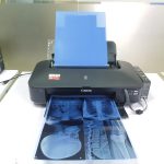
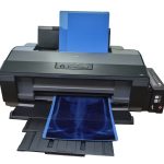
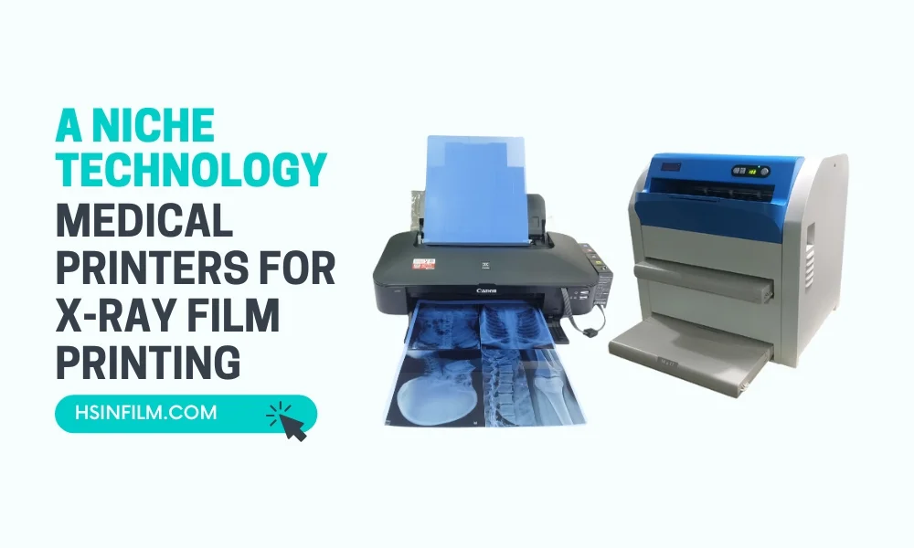
Also read: Applications of Medical Thermal Printers
Types of Orthodontic X-Rays and When They’re Used
Panoramic X-Rays
Panoramic X-rays capture the entire mouth in a single image, including the teeth, upper and lower jaws, and surrounding structures.
- Advantages: Provide a comprehensive view of the mouth, helping to identify impacted teeth, jaw issues, and other abnormalities.
- Usage: Commonly used during the initial examination and throughout treatment to monitor progress.
Cephalometric X-Rays
Cephalometric X-rays focus on the side profile of the head, showing the relationship between the teeth, jaw, and skull.
- Advantages: Helps in understanding the growth patterns of the jaw and planning treatments that address facial structure and bite alignment.
- Usage: Essential for diagnosing and planning treatments for orthodontic issues related to jaw alignment and facial growth.
Periapical X-Rays
Periapical X-rays provide detailed images of one or two teeth, including the root and surrounding bone.
- Advantages: Useful for identifying issues with individual teeth, such as infections, cysts, or other abnormalities.
- Usage: Often used to diagnose specific problems or to monitor the health of individual teeth during treatment.
Bitewing X-Rays
Bitewing X-rays show the upper and lower teeth in a specific area of the mouth, highlighting the crowns of the teeth and the height of the bone between them.
- Advantages: Useful for detecting decay between teeth and monitoring changes in bone density.
- Usage: Commonly used in routine dental check-ups and during orthodontic treatment to check for decay and bone health.
How Orthodontic X-Rays Are Taken
Preparation
Before taking an X-ray, the orthodontist or dental technician will explain the procedure and position the patient appropriately. The patient may be asked to wear a lead apron to protect against radiation.
Procedure
The procedure for taking an orthodontic X-ray is quick and painless:
- Positioning: The patient is positioned correctly, either sitting or standing, depending on the type of X-ray.
- Exposure: The X-ray machine is positioned, and the patient is asked to remain still while the image is taken.
- Image Capture: The X-ray image is captured in a matter of seconds, and the patient can resume normal activities immediately afterward.
Safety Considerations
Orthodontic X-rays involve minimal radiation exposure, but safety measures are always taken to protect patients. Lead aprons and collars are used to shield the body from unnecessary exposure, and modern X-ray machines are designed to minimize radiation doses.
Interpreting Orthodontic X-Rays
Reading the Images
Orthodontists are trained to read and interpret X-ray images, identifying any issues and determining the best course of treatment. Common findings include misaligned teeth, impacted teeth, jaw abnormalities, and signs of decay or infection.
Diagnosing Issues
Based on the X-ray images, the orthodontist can diagnose a variety of issues:
- Misaligned Teeth: Identifying teeth that are out of alignment or crowded.
- Impacted Teeth: Detecting teeth that have not erupted properly.
- Jaw Issues: Diagnosing problems with the jaw structure or alignment.
- Decay and Infection: Spotting signs of decay, cysts, or infections that need treatment.
Role of X-Rays in Orthodontic Treatment

Treatment Planning
X-rays are crucial in planning orthodontic treatments. They provide detailed information about the position and condition of the teeth and jaw, allowing the orthodontist to develop a customized treatment plan.
- Braces: Determining the need for braces and planning the placement of brackets and wires.
- Extractions: Identifying teeth that may need to be extracted to make space for proper alignment.
- Surgical Interventions: Planning any necessary surgical procedures to correct jaw alignment or remove impacted teeth.
Monitoring Progress
Throughout orthodontic treatment, X-rays are used to monitor progress and make adjustments as needed. Regular X-rays help ensure that the treatment is moving in the right direction and allow the orthodontist to make any necessary changes to the treatment plan.
Benefits of Orthodontic X-Rays
Accurate Diagnosis
X-rays provide a clear and detailed view of the teeth and jaw, enabling accurate diagnosis of orthodontic issues that may not be visible during a regular dental exam.
Personalized Treatment
With the detailed information provided by X-rays, orthodontists can develop personalized treatment plans that address the specific needs of each patient.
Improved Outcomes
Regular monitoring with X-rays ensures that orthodontic treatments are progressing as planned, leading to better outcomes and a healthier smile.
Comparing Traditional and Digital Orthodontic X-rays
Pros and Cons Unveiled
Traditional film X-rays have been a staple, but digital X-rays have stepped into the spotlight. Digital technology offers instant results, reduced environmental impact, and enhanced image quality. It’s akin to switching from a classic film camera to the latest smartphone camera—embracing efficiency and innovation.
Digital Impact on Orthodontic Diagnostics
Digital X-rays have transformed the way orthodontic diagnostics are conducted. They allow for easy storage, retrieval, and sharing of images, fostering collaborative treatment planning. It’s like upgrading from a filing cabinet to a cloud-based system—streamlining processes for better patient care.
Orthodontic X-Rays for Early Intervention
Early Detection, Lifelong Benefits
X-rays aren’t just for adults; they are superheroes in identifying orthodontic issues in children. Early intervention based on these images ensures timely correction, preventing potential complications down the road. It’s like planting a seed for a healthy smile that will bloom over the years.
Adult Orthodontics
Tailoring Treatment for Adults
X-rays are equally vital in adult orthodontics, helping orthodontists navigate the unique challenges presented by mature jaws and teeth. It’s like adjusting the course of a ship—navigating the seas of orthodontic challenges with precision.
Addressing Specific Challenges
Adults might face issues like tooth wear, bone loss, and gum disease. Orthodontic X-rays serve as the compass, guiding professionals in creating effective treatment plans that address these challenges head-on.
Conclusion
Orthodontic X-rays are an essential tool in modern dental care, providing valuable information for diagnosing and treating a wide range of orthodontic issues. Understanding the different types of X-rays, how they are taken, and their role in treatment can help patients feel more comfortable and informed about their orthodontic care.
By providing accurate and detailed images, X-rays enable orthodontists to develop personalized treatment plans, monitor progress, and ensure the best possible outcomes for their patients. Whether you’re starting your orthodontic journey or are already in treatment, knowing the importance and benefits of X-rays can help you appreciate the care and precision that goes into creating a beautiful, healthy smile.
Frequently Asked Questions about Orthodontic X-Rays
1. Are orthodontic x-rays safe?
Absolutely. Orthodontic x-rays follow strict safety measures, including protective gear and advanced technology, ensuring your safety comes first.
2. How often do I need orthodontic x-rays?
The frequency depends on your individual case, but generally, x-rays are taken at the beginning of treatment and periodically to monitor progress.
3. Do x-rays hurt?
Not at all. Taking orthodontic x-rays is painless. You might feel a slight discomfort, but it’s quick and harmless.
4. Are digital x-rays better than traditional ones?
Digital x-rays offer advantages like speed and clarity, but both types are effective. Your orthodontist will determine the best approach for your specific needs.
5. Why do children need orthodontic x-rays?
Early intervention is key to addressing potential issues. X-rays help identify and correct problems, ensuring a lifetime of healthy smiles.