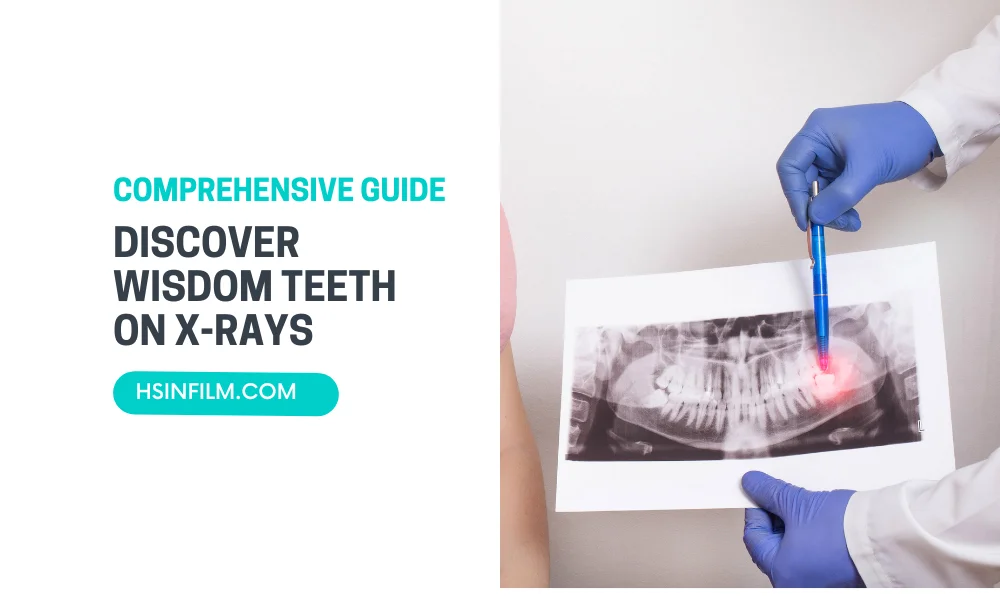
Wisdom teeth, also known as third molars, are the last set of teeth to emerge in the back corners of your mouth. They usually appear between the ages of 17 and 25. Sometimes, these teeth can cause problems, such as pain, infection, or crowding, which is why it’s important to know how to identify them and understand their development. One of the best ways to do this is through X-rays. This blog post will guide you on what to expect when discovering wisdom teeth on X-rays, why they are important, and what the process involves.
Table of Contents
Understanding Wisdom Teeth
What Are Wisdom Teeth?
Wisdom teeth are the third set of molars located at the very back of your mouth. They are the last teeth to develop and can be found in the upper and lower jaws. Most people have four wisdom teeth, but it’s not uncommon to have fewer or even none at all.
Why Are They Called Wisdom Teeth?
The name “wisdom teeth” comes from the age at which they typically appear. They emerge during the late teenage years or early twenties, a time traditionally associated with gaining maturity and “wisdom.”
Also read: Understanding Orthodontic X-ray
The Importance of X-Rays for Wisdom Teeth
Why Use X-Rays?
X-rays are crucial for identifying the position and development of wisdom teeth. They provide a clear image of the teeth and jawbone, helping dentists determine if the wisdom teeth are coming in properly or if there are any issues.
What Can X-Rays Reveal?
X-rays can show the exact location of wisdom teeth, their orientation, and whether they are impacted (trapped under the gum or jawbone). They can also reveal any potential problems, such as crowding, cysts, or damage to nearby teeth.
Types of X-Rays Used
Panoramic X-Rays
Panoramic X-rays capture a broad view of the entire mouth, including all the teeth, jawbone, and surrounding structures. This type of X-ray is commonly used to assess the development and position of wisdom teeth.
Periapical X-Rays
Periapical X-rays focus on a smaller area and provide detailed images of one or two teeth, including their roots and surrounding bone. These are useful for examining specific teeth or areas of concern.
Cone Beam CT Scans
Cone beam CT scans provide 3D images of the teeth, bones, and soft tissues. They offer a detailed view and are particularly useful in complex cases where a more comprehensive assessment is needed.
How X-Rays Track Wisdom Teeth Development
Wisdom teeth, the last set of molars that typically emerge in late adolescence or early adulthood, often bring questions about their development and impact on oral health. X-rays play a crucial role in tracking the journey of these teeth. This section explores the specific ways in which X-rays act as a reliable guide in monitoring and understanding the development of wisdom teeth.
1. Visualizing Formation:
X-rays provide a clear view of the initial stages of wisdom teeth formation. Through detailed imaging, dentists can observe the growth process, ensuring that teeth develop properly in alignment with the jaw.
2. Monitoring Eruption:
As wisdom teeth begin to emerge, X-rays track their eruption into the oral cavity. This dynamic imaging allows dental professionals to monitor the timing and positioning of the teeth, identifying any potential issues that may arise during this process.
3. Assessing Alignment:
One of the critical aspects of wisdom teeth development is their alignment with existing teeth. X-rays offer a comprehensive view of the entire dental landscape, aiding dentists in assessing whether the emerging teeth are positioned correctly or if adjustments are needed.
4. Detecting Impaction:
X-rays are particularly effective in detecting impaction, a common occurrence where wisdom teeth do not fully emerge. By visualizing the extent of impaction, dentists can determine the appropriate course of action, whether it be monitoring or recommending extraction.
5. Noting Complications:
Complications such as overcrowding or misalignment can be observed through X-rays. This detailed imaging allows dental professionals to identify potential challenges and proactively address them to maintain overall oral health.
Navigating Wisdom Teeth Challenges: Impaction and Overcrowding Unveiled in X-Rays
Exploring the world of wisdom teeth reveals common hurdles—impaction and overcrowding. Through the lens of X-rays, we navigate these challenges, deciphering their relevance to the unique journey of wisdom teeth and understanding their impact on oral health.
1. Unmasking Impaction:
X-rays serve as a revealing tool in unmasking impaction, where wisdom teeth struggle to emerge fully. Precise imaging allows dentists to assess the extent of impaction, guiding decisions on monitoring or extraction. This insight is crucial, preventing potential complications like pain and infections associated with impacted wisdom teeth.
2. Decoding Overcrowding Dilemmas:
Overcrowding, captured vividly in X-rays, occurs when there’s limited space for wisdom teeth to comfortably emerge. These images provide a detailed understanding of overcrowding’s impact on existing teeth alignment. Deciphering this dilemma early on enables proactive measures to maintain proper dental harmony.
3. Wisdom Teeth’s Unique Significance:
The relevance of these concerns is intricately tied to the uniqueness of wisdom teeth. Impaction and overcrowding can disrupt the natural development of these latecomers, impacting the overall oral landscape. Recognizing their significance in the context of wisdom teeth underscores the need for attentive monitoring and tailored interventions.
4. Oral Health Implications:
Beyond their impact on wisdom teeth, these challenges bear direct consequences for oral health. Impacted teeth may lead to discomfort and infections, while overcrowding poses risks of misalignment. X-rays play a pivotal role in uncovering these issues, allowing for timely interventions that safeguard both the integrity of wisdom teeth and the overall oral well-being.
Signs of Wisdom Teeth Troubles in X-Rays
Embarking on a diagnostic journey through X-rays, this section unveils the visual cues that indicate potential troubles associated with wisdom teeth. X-ray images serve as powerful tools, allowing dentists to identify common problems that may lead to discomfort and dental complications. Join us as we decode the signs captured in these images, shedding light on the issues that may lurk beneath the surface.
1. Identifying Impacted Wisdom Teeth:
X-rays are instrumental in identifying signs of impacted wisdom teeth, where the molars struggle to fully emerge. Through detailed imaging, we explore the indicators of impaction, discerning whether the teeth are partially or completely obstructed. Uncover the visual language that reveals the presence of impacted wisdom teeth.
2. Spotting Misalignment:
Misalignment, a potential troublemaker, is highlighted in X-rays. These images provide a clear view of how wisdom teeth may emerge at angles that disrupt the natural alignment of adjacent teeth. Dive into the visuals to spot signs of misalignment and understand its implications on overall dental harmony.
3. Uncovering Potential Complications:
Complications such as infections or cysts can be lurking beneath the surface, and X-rays are the key to uncovering these hidden threats. This section delves into the signs within the images that may indicate the presence of complications, emphasizing the importance of proactive measures to prevent and manage potential issues.
4. Decoding Discomfort Indicators:
Discomfort associated with wisdom teeth troubles can be visualized through X-rays. Explore how these images capture subtle indicators of discomfort, guiding dentists in understanding the root cause. By decoding these signs, dental professionals can tailor appropriate interventions for optimal patient comfort.
Connecting the Dots: From X-Ray to Dental Complications
1. Unveiling the Hidden: X-Ray as a Diagnostic Tool
X-rays play a pivotal role in unveiling hidden aspects of oral health. X-rays act as the starting point, revealing the canvas upon which potential dental complications are painted.
2. Identifying Complications through Visual Clues
With the aid of X-rays, dentists can identify visual clues that point towards potential complications. By connecting these visual dots, dental professionals can initiate targeted interventions to address emerging problems.
3. Proactive Intervention: From X-Ray Insights to Treatment Plans
X-rays not only identify complications but also pave the way for proactive intervention. By connecting the dots between X-ray findings and potential complications, dentists can tailor interventions that address issues at their roots, ensuring effective and targeted care.
4. Preventive Measures: Mitigating Future Complications
The journey concludes by emphasizing the role of X-rays in preventive dentistry. Through early detection of potential complications, X-rays empower dental professionals to implement preventive measures. This proactive approach mitigates the risk of future dental issues, creating a pathway towards long-term oral health.

Proactive Wisdom: The Importance of X-Ray Assessment
In this pivotal section, we delve into the proactive realm of wisdom teeth care, emphasizing the crucial role of X-ray assessment as a preemptive tool. By harnessing the power of X-rays before symptoms emerge, individuals and dental professionals can assess wisdom teeth health proactively. This proactive approach, especially beneficial in younger individuals, offers a spectrum of advantages, allowing potential issues to be identified and addressed before they have the chance to escalate.
1. Anticipating Potential Issues:
The proactive use of X-rays allows for the anticipation of potential issues before they manifest as symptoms. By gaining insight into the development and alignment of wisdom teeth through early assessments, dental professionals can foresee and address issues, mitigating the risk of discomfort or complications.
2. Tailoring Preventive Strategies:
Early X-ray assessments enable the tailoring of preventive strategies. Identifying subtle signs of misalignment or impaction empowers dental professionals to devise personalized plans that may include monitoring, early interventions, or recommendations for optimal oral health outcomes.
3. Empowering Younger Individuals:
The benefits of proactive X-ray assessment are particularly pronounced in younger individuals. By initiating assessments at an early age, dental professionals can guide the proper development of wisdom teeth, addressing issues during their formative stages. This proactive approach lays the foundation for a healthier oral landscape in adulthood.
4. Minimizing Future Complications:
Proactive wisdom through X-ray assessment minimizes the risk of future complications. By identifying and addressing potential issues early on, individuals can avoid the discomfort and disruptions that may arise if problems are left unattended. This proactive stance contributes to overall oral well-being and a smoother oral health journey.
Step-by-Step: Analyzing Wisdom Teeth on X-Rays
Behind the Scenes: How Dentists Analyze X-Rays
Embark on a journey behind the scenes of the dental office, where the intricate process of analyzing X-rays unfolds. This section unveils the meticulous steps and expert insights that dentists employ to decode the information captured in X-ray images, offering a glimpse into the fascinating world of diagnostic precision.
1. Capturing the Images:
The process begins with the capturing of X-ray images, a step-by-step procedure where dental professionals ensure the clarity and accuracy of each shot. This section outlines the technology and techniques involved in obtaining high-quality images that serve as the foundation for insightful analysis.
2. Image Processing:
Once the images are captured, they undergo a meticulous processing phase. Explore how advanced technology aids in enhancing and refining the X-ray images. This step is crucial in ensuring that the details necessary for accurate diagnosis are crystal clear.
3. Interpreting the Visual Landscape:
Dentists then step into the role of visual interpreters. This part of the journey delves into how dental professionals analyze the visual landscape of X-rays. From assessing tooth development to identifying potential complications, dentists use their expertise to decode the intricate details embedded in the images.
4. Formulating a Diagnosis:
The culmination of the process lies in formulating a diagnosis. Discover how dentists connect the dots, correlating the visual information from X-rays with clinical observations and patient history. This section emphasizes the diagnostic precision that X-ray analysis brings to the forefront.
5. Communication with Patients:
The journey concludes with communication. Dentists take the insights gained from X-ray analysis and translate them into clear, comprehensible explanations for patients. Explore how effective communication bridges the gap between complex diagnostic processes and the understanding of individuals seeking dental care.
To Extract or Not: Deciphering the Wisdom Teeth X-Ray Code
In the realm of wisdom teeth, the decision to extract or preserve hinges on a thorough deciphering of the X-ray code. This section unravels the complexities surrounding wisdom teeth X-rays, providing insight into how dental professionals navigate the visual language to make informed decisions about extraction or preservation.
1. Interpreting Positioning and Alignment:
Wisdom teeth X-rays unveil the positioning and alignment of these molars. This part of the journey explores how dental professionals interpret the angles and placement of wisdom teeth to determine whether they are in harmony with the existing dental landscape or if misalignment and potential issues are evident.
2. Assessing Impaction Levels:
A key aspect of the X-ray code lies in assessing the degree of impaction. Delve into how dentists use X-ray images to identify whether wisdom teeth are partially or completely impacted. This critical information guides decisions on whether extraction is necessary to prevent complications associated with impaction.
3. Gauging Developmental Stage:
Wisdom teeth X-rays also serve as a timeline, capturing the developmental stages of these molars. Explore how dental professionals gauge whether the wisdom teeth are fully developed or still in the process of emerging. This knowledge influences decisions on whether extraction is timely or if monitoring is a viable option.
4. Identifying Complications:
Complications, visible in X-ray images, play a significant role in the decision-making process. Uncover how dental professionals identify signs of potential issues such as infections, cysts, or damage to adjacent teeth. This section sheds light on how the presence of complications may tip the scale toward extraction.
5. Balancing Individual Factors:
The X-ray code is not one-size-fits-all. This part of the journey explores how dental professionals balance the individual factors of each patient, considering aspects like age, overall oral health, and the potential impact of wisdom teeth on the patient’s well-being. This personalized approach ensures decisions align with the unique needs of each individual.
Conclusion
Understanding what to expect when discovering wisdom teeth on X-rays is crucial for maintaining good oral health. X-rays provide valuable insights into the development and position of wisdom teeth, allowing for early detection and treatment of potential issues. Regular dental check-ups, good oral hygiene, and a healthy diet are essential preventive measures. If your wisdom teeth are causing problems, consult with your dentist to determine the best course of action. By staying informed and proactive, you can ensure that your wisdom teeth do not disrupt your oral health.
HSIN Film’s Contribution to Your Dental Health
Discover how HSIN Film’s medical dry film, thermal film, laser film, thermal printer, and laser printer enhance the clarity of wisdom teeth X-rays. With cutting-edge technology, HSIN Film ensures that every detail is captured for an accurate diagnosis.


