Technology stands as a cornerstone in elevating diagnostics and patient care in modern dentistry. A standout among these innovations is the Phosphor Plate (PSP) system, a technological marvel reshaping dental radiography. This comprehensive guide unravels the intricacies of Phosphor Plates in Dental X-rays, delving into their technology, advantages, workflow, and best practices, offering a transformative approach to mastering dental X-rays effectively.
Table of Contents
Understanding Phosphor Plates
What are Phosphor Plates?
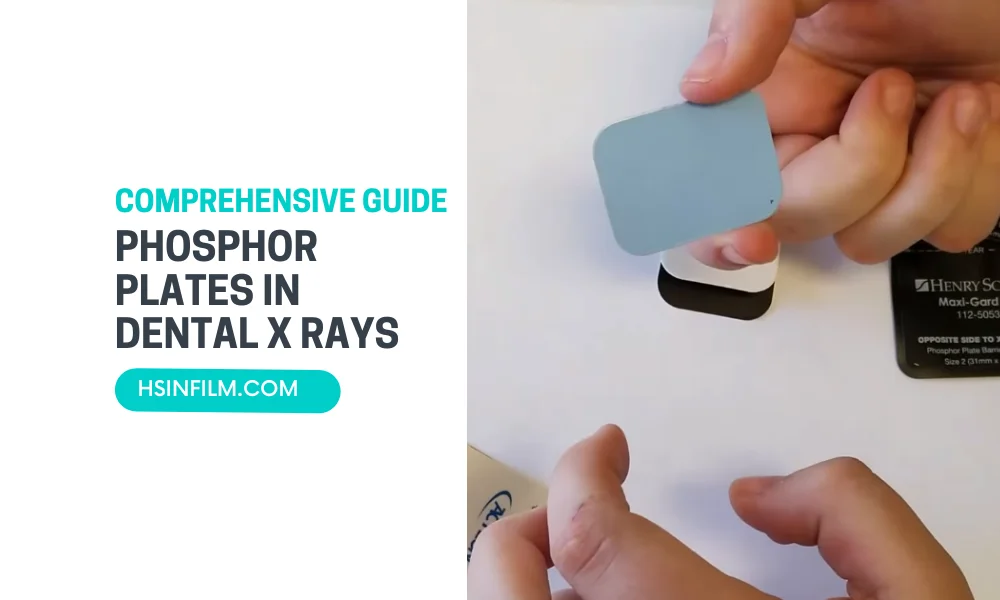
Phosphor Plates are flexible, wireless alternatives to traditional film-based X-rays. They are coated with a photostimulable phosphor layer that captures X-ray energy during exposure.
How Do They Work?
During an X-ray exposure, the phosphor layer absorbs X-ray photons. After exposure, the plates are scanned with a laser, releasing the stored energy as light. This light is then converted into a digital image.
Specific Applications of Phosphor Plates in Different Dental Procedures
Phosphor plates, also known as photostimulable phosphor (PSP) plates, are a type of digital imaging technology used in dental radiography. They play a crucial role in various dental procedures due to their flexibility, ease of use, and ability to produce high-quality images. Here’s a detailed exploration of their specific applications in different dental procedures:
1. Diagnostic Imaging
- Routine Dental Exams: Phosphor plates are commonly used for routine dental check-ups to capture detailed images of the teeth, bones, and surrounding structures. This helps in identifying cavities, bone loss, and other dental issues that may not be visible during a physical examination.
- Caries Detection: Phosphor plates are highly sensitive, making them excellent for detecting early-stage caries (tooth decay). The high resolution of the images allows dentists to spot even small areas of demineralization.
2. Endodontic Procedures
- Root Canal Therapy: During root canal procedures, phosphor plates are used to take periapical X-rays, which show the entire tooth from the crown to the tip of the root. These images are crucial for diagnosing the extent of infection, planning the treatment, and monitoring the progress of the procedure.
- Pulp Vitality Testing: Phosphor plates can be used to capture images that help assess the vitality of the tooth pulp, aiding in decisions about whether a root canal is necessary.
3. Periodontal Assessment
- Bone Level Evaluation: In periodontal treatment, phosphor plates are used to assess the bone levels around teeth, which is important for diagnosing periodontal disease. The high-resolution images help in evaluating the extent of bone loss and planning appropriate treatments.
- Monitoring Bone Regeneration: After periodontal surgeries, phosphor plates can be used to monitor bone regeneration over time, providing detailed images that help assess the effectiveness of the treatment.
4. Oral Surgery
- Impacted Tooth Assessment: Phosphor plates are used to capture detailed images of impacted teeth, such as wisdom teeth. These images are critical for planning surgical extractions, as they provide a clear view of the tooth’s position relative to the surrounding structures.
- Pre-surgical Planning: For other oral surgeries, such as implant placement or cyst removal, phosphor plates provide high-quality images that help in planning the procedure and avoiding complications.
5. Orthodontics
- Treatment Planning: Phosphor plates are often used in orthodontic assessments to capture images of the teeth and jaw, which are essential for planning treatments like braces or aligners. The detailed images help orthodontists assess the alignment of teeth and the growth patterns of the jaw.
- Progress Monitoring: Throughout orthodontic treatment, phosphor plates can be used to take periodic images to monitor the movement of teeth and adjust the treatment plan as needed.
6. Prosthodontics
- Crown and Bridge Placement: Phosphor plates are used to capture images before and after the placement of crowns and bridges. These images help in evaluating the fit and alignment of the prosthetics.
- Implant Planning: When planning for dental implants, phosphor plates provide detailed images of the jawbone, allowing for precise placement of the implants and reducing the risk of complications.
7. Pediatric Dentistry
- Growth and Development Monitoring: In pediatric dentistry, phosphor plates are used to monitor the development of primary and permanent teeth. These images help in identifying any abnormalities or delays in tooth eruption.
- Trauma Assessment: Phosphor plates are also used to assess dental trauma in children, such as fractured teeth or injuries to the jaw, providing clear images that aid in diagnosis and treatment planning.
Advantages of Phosphor Plates in Dental Procedures
- High Image Quality: Phosphor plates produce high-resolution images that are crucial for accurate diagnosis and treatment planning.
- Flexible and Reusable: The plates are flexible and can be reused multiple times, making them cost-effective and patient-friendly.
- Wide Dynamic Range: Phosphor plates can capture a wide range of densities, from soft tissues to hard bones, providing comprehensive images for various dental procedures.
Disadvantages of Phosphor Plates
- Image Processing Time: Unlike direct digital sensors, phosphor plates require a separate scanning step to process the images, which can be time-consuming.
- Handling Sensitivity: Phosphor plates are more sensitive to scratches and light exposure, which can affect image quality if not handled properly.
Phosphor plates are a versatile and valuable tool in dental radiography, playing a critical role in a wide range of dental procedures. Their ability to provide detailed, high-quality images makes them indispensable for accurate diagnosis, effective treatment planning, and ongoing patient care.
Also read: How to build an X-Ray Room?
Compare and Contrast Phosphor Plates with Other Digital Imaging Technologies
Phosphor plates (PSP) are one of several digital imaging technologies used in dental radiography. To understand their unique advantages and limitations, it’s useful to compare them with other common digital imaging technologies like digital sensors (both CCD and CMOS) and cone beam computed tomography (CBCT). Here’s a detailed comparison:
1. Image Quality
- Phosphor Plates (PSP)
- Advantages: PSP plates offer high-resolution images with a wide dynamic range, capturing fine details in both hard and soft tissues. This makes them suitable for various dental procedures, from routine check-ups to complex endodontic treatments.
- Limitations: While PSP plates provide excellent image quality, the resolution can sometimes be lower than that of direct digital sensors, especially in very fine details.
- Digital Sensors (CCD/CMOS)
- Advantages: CCD (Charge-Coupled Device) and CMOS (Complementary Metal-Oxide-Semiconductor) sensors typically provide superior image quality, with higher resolution and sharper images. They are particularly good for detailed diagnostics and are immediately available after exposure.
- Limitations: The sensors are rigid and can be uncomfortable for patients, especially in areas that are difficult to reach.
- CBCT
- Advantages: CBCT provides 3D imaging, offering detailed views of anatomical structures that are not possible with 2D imaging. It is particularly useful for complex cases like implant planning, bone assessment, and detailed evaluation of anatomical relationships.
- Limitations: CBCT exposes patients to higher radiation doses and is more expensive. The technology also requires more extensive training to interpret the images correctly.
2. Ease of Use and Patient Comfort
- Phosphor Plates (PSP)
- Advantages: PSP plates are flexible and thinner than digital sensors, making them more comfortable for patients, particularly in pediatric dentistry or when imaging hard-to-reach areas.
- Limitations: The process of scanning the plates after exposure can be time-consuming, as it requires a separate step before the image is available.
- Digital Sensors (CCD/CMOS)
- Advantages: Digital sensors provide immediate image availability, streamlining the diagnostic process and allowing for real-time adjustments.
- Limitations: The rigidity and thickness of these sensors can cause discomfort, especially for patients with small mouths or gag reflexes.
- CBCT
- Advantages: CBCT scans are non-invasive and relatively quick, often more comfortable than multiple traditional X-rays for complex cases.
- Limitations: The patient must remain very still during the scan, which can be difficult for some patients, particularly children.
3. Workflow Efficiency
- Phosphor Plates (PSP)
- Advantages: PSP systems are generally easy to integrate into existing workflows and can be used with most traditional X-ray equipment, making them a good choice for practices transitioning from film to digital.
- Limitations: The additional step of scanning the plate before viewing the image can slow down the workflow compared to direct digital sensors.
- Digital Sensors (CCD/CMOS)
- Advantages: These sensors streamline the workflow by providing instant images, which can be viewed and analyzed immediately, allowing for quick decisions during procedures.
- Limitations: The initial cost of the sensors can be high, and they may require specialized equipment, adding complexity to the setup.
- CBCT
- Advantages: CBCT is efficient for complex cases, reducing the need for multiple 2D images and providing comprehensive information in a single scan.
- Limitations: The setup and interpretation are more complex, and the higher cost may limit its routine use in some dental practices.
4. Cost and Maintenance
- Phosphor Plates (PSP)
- Advantages: PSP systems are generally more affordable than digital sensors and CBCT. The plates are reusable, which can reduce long-term costs.
- Limitations: PSP plates are sensitive to scratches, dust, and light exposure, which can degrade image quality over time, requiring careful handling and maintenance.
- Digital Sensors (CCD/CMOS)
- Advantages: While the upfront cost is higher, digital sensors typically have a long lifespan and require minimal ongoing maintenance.
- Limitations: The high initial investment and the potential for damage due to drops or bites can be costly.
- CBCT
- Advantages: CBCT is a powerful diagnostic tool, but its high cost and maintenance requirements limit its use to larger practices or specific cases.
- Limitations: The equipment is expensive, and the ongoing maintenance can be significant, making it less accessible for smaller practices.
5. Radiation Exposure
- Phosphor Plates (PSP)
- Advantages: PSP systems generally require lower radiation doses than traditional film but may require slightly higher doses compared to direct digital sensors.
- Limitations: The radiation dose can vary depending on the type of imaging and the specific equipment used.
- Digital Sensors (CCD/CMOS)
- Advantages: These sensors are highly sensitive, often requiring the lowest radiation doses among all digital imaging options, enhancing patient safety.
- Limitations: There is a trade-off between image quality and radiation dose, with very low doses sometimes leading to less detailed images.
- CBCT
- Advantages: While providing more comprehensive information, CBCT generally involves higher radiation exposure compared to 2D imaging methods. However, it can be more efficient in reducing overall radiation when replacing multiple 2D images.
- Limitations: The higher dose can be a concern, particularly in cases where CBCT may not be strictly necessary.
Phosphor plates (PSP) offer a balanced approach between image quality, patient comfort, and cost-effectiveness, making them a versatile option in many dental practices. However, they require careful handling and an extra processing step, which may not suit every practice. Digital sensors (CCD/CMOS) provide superior image quality and immediate results, but at a higher cost and with potential patient comfort issues. CBCT stands out for complex cases requiring detailed 3D imaging, though it comes with higher costs and radiation exposure.
Choosing the right digital imaging technology depends on the specific needs of the practice, the types of procedures performed, and the priorities in terms of image quality, workflow efficiency, patient comfort, and cost.
Role of Image Processing Algorithms in Optimizing Phosphor Plate Images and Improving Diagnostic Accuracy
Image processing algorithms play a crucial role in optimizing Phosphor Plate (PSP) images, enhancing their quality, and improving diagnostic accuracy in dental radiography. Phosphor Plates capture X-ray images by converting them into digital form, but the raw images often require post-processing to achieve the best diagnostic outcomes. Here’s how image processing algorithms contribute to this process:
1. Noise Reduction
- Role: Noise, which appears as random variations in brightness or color, can obscure fine details in an X-ray image, leading to diagnostic errors. Image processing algorithms are designed to reduce noise while preserving critical information.
- Techniques:
- Median Filtering: Smooths the image by replacing each pixel’s value with the median value of the surrounding pixels. This technique is effective in reducing ‘salt-and-pepper’ noise without blurring edges.
- Gaussian Filtering: Applies a Gaussian function to smooth the image, reducing noise while maintaining important structural details.
2. Contrast Enhancement
- Role: Contrast enhancement algorithms improve the visibility of anatomical structures in PSP images by making the differences between various tissues more pronounced. This is particularly important in dental imaging, where subtle differences in tissue density can indicate conditions like caries or bone loss.
- Techniques:
- Histogram Equalization: Redistributes the intensity values of the image to cover the full range of possible values, resulting in better contrast.
- Adaptive Contrast Enhancement: Adjusts contrast differently across various regions of the image, enhancing areas of interest without overexposing other regions.
3. Edge Detection and Enhancement
- Role: Edge detection algorithms are critical for identifying the boundaries of structures such as teeth, roots, and lesions. Enhanced edge detection helps in accurately outlining these structures, which is vital for diagnosis and treatment planning.
- Techniques:
- Sobel and Canny Edge Detectors: These are popular methods for detecting edges in images. They work by identifying areas where there is a significant change in intensity, which usually corresponds to the edges of structures.
- Unsharp Masking: Enhances edges by subtracting a blurred version of the image from the original image, making the edges stand out more clearly.
4. Dynamic Range Adjustment
- Role: PSP images have a wide dynamic range, meaning they can capture a broad range of intensity levels. However, not all parts of the image may be within the optimal viewing range. Dynamic range adjustment algorithms help in compressing or expanding this range to make all parts of the image visible and diagnostically useful.
- Techniques:
- Logarithmic and Exponential Transformations: Apply mathematical functions to adjust the dynamic range, improving visibility in darker or lighter regions of the image.
- Gamma Correction: Adjusts the brightness of the image by applying a gamma function, which can either brighten or darken the image depending on the needs of the diagnosis.
5. Image Sharpening
- Role: Image sharpening algorithms enhance the clarity and detail of PSP images, making it easier to identify small or subtle features that could indicate pathology.
- Techniques:
- High-Pass Filtering: Enhances fine details by amplifying high-frequency components, which correspond to edges and fine structures in the image.
- Laplacian Filtering: Emphasizes regions of rapid intensity change, making edges and fine details more prominent.
6. Artifact Removal
- Role: PSP images can sometimes contain artifacts—unwanted elements that are not part of the actual image, such as scratches, dust marks, or scanner defects. Artifact removal algorithms are designed to eliminate or reduce these artifacts to avoid misinterpretation.
- Techniques:
- Morphological Operations: Used to remove small artifacts by applying operations like erosion and dilation, which can effectively clean up the image.
- Inpainting Algorithms: Replace the area of the artifact with a patch of the image that matches the surrounding area, effectively ‘painting over’ the unwanted elements.
7. Image Registration and Alignment
- Role: In some dental procedures, multiple images may be taken from different angles or at different times. Image registration algorithms align these images so they can be compared or combined, improving the diagnostic process.
- Techniques:
- Rigid and Non-Rigid Registration: Aligns images by translating, rotating, or deforming them to match, allowing for accurate comparison or integration of multiple images.
- Cross-Correlation: A method for finding the best alignment between two images by shifting one image relative to the other and finding the position that maximizes their similarity.
8. Automatic Segmentation
- Role: Segmentation algorithms divide the image into regions corresponding to different anatomical structures, such as teeth, bones, or soft tissues. This automatic division helps in focusing on specific areas of interest for more detailed analysis.
- Techniques:
- Thresholding: Divides the image based on intensity values, useful for separating hard tissues from soft tissues.
- Region Growing: Starts from a seed point and expands the region by adding neighboring pixels that have similar properties, useful for identifying specific structures.
9. Quantitative Analysis
- Role: Image processing algorithms also enable quantitative analysis of PSP images, such as measuring distances, angles, and areas. This is important for treatment planning and monitoring the progression of conditions.
- Techniques:
- Automated Measurement Tools: Algorithms can automatically measure structures in the image, providing precise data for diagnosis and treatment.
- 3D Reconstruction: In some advanced cases, algorithms can be used to reconstruct 3D models from 2D images, offering a more comprehensive view of the anatomy.
Image processing algorithms are essential in optimizing Phosphor Plate images for diagnostic use in dental practices. By enhancing image quality, reducing noise, improving contrast, and automatically identifying anatomical structures, these algorithms significantly improve the accuracy and reliability of diagnoses. As dental imaging technology continues to advance, the role of sophisticated image processing will only become more critical, helping dentists to deliver better patient care through more accurate and detailed radiographic evaluations.
Workflow with Phosphor Plates
Image Acquisition
The dentist places the Phosphor Plate inside the patient’s mouth to capture the X-ray image. The thin, flexible nature of the plates ensures a comfortable experience for the patient.
Plate Scanning
Once exposed, the plates are removed and scanned using a dedicated imaging system. The laser scanner extracts the stored energy, producing a digital image ready for analysis.
Digital Image Viewing
The digital images are instantly available for viewing on computer screens. Dentists can zoom in, adjust contrast, and apply various filters to enhance diagnostic capabilities.
Best Practices for Using Phosphor Plates
Proper Handling
Handle Phosphor Plates with care to avoid scratches or damage. Always use protective barriers to maintain hygiene standards.
Regular Maintenance
Ensure the laser scanner is well-maintained and calibrated regularly to guarantee accurate and clear digital images.
Patient Communication
Explain the advantages of Phosphor Plates to patients, emphasizing the reduced radiation exposure and enhanced comfort during X-ray procedures.
Training and Education
Dental professionals should undergo training on the proper use of Phosphor Plates, ensuring optimal image quality and diagnostic accuracy.

Mastering Phosphor Plates in Dental X-Rays Effectively: A Step-by-Step Guide
In the dynamic field of dentistry, staying at the forefront of technological advancements is key to providing optimal patient care. One such innovation that has revolutionized dental imaging is the use of Phosphor Plates in Dental X-rays. This guide aims to take you through a step-by-step journey, unlocking the secrets to mastering dental X-rays with Phosphor Plates effectively.
Step 1: Understanding the Technology
What Are Phosphor Plates?
Phosphor Plates are flexible, reusable imaging plates coated with a phosphor layer. This layer stores X-ray energy during exposure, which is later released and converted into a digital image during the scanning process.
How Do They Work?
When exposed to X-rays, the phosphor layer in the plate absorbs energy. After exposure, the plate is scanned using a laser, releasing the stored energy as light. This light is then transformed into a digital image, offering a detailed view of the oral structures.
Step 2: Advantages of Phosphor Plates in Dental X-rays
Enhanced Diagnostic Capabilities
Phosphor Plates deliver high-resolution images, capturing fine details crucial for accurate diagnostics. The digital format allows for easy manipulation, enhancing diagnostic capabilities.
Patient Comfort
The flexibility and thinness of Phosphor Plates ensure a more comfortable experience for patients during intraoral X-rays. They adapt to the contours of the oral cavity, reducing discomfort.
Reduced Radiation Exposure
Compared to traditional film X-rays, Phosphor Plates require less radiation for image acquisition. This not only enhances patient safety but also aligns with the principles of ALARA (As Low As Reasonably Achievable) radiation exposure.
Step 3: Workflow with Phosphor Plates
Image Acquisition
The dentist places the Phosphor Plate in the patient’s mouth to capture the X-ray image. The flexibility of the plate ensures it conforms comfortably to the oral anatomy.
Plate Scanning
After exposure, the plate is removed and placed in a dedicated laser scanner. The scanning process releases the stored energy, creating a digital image that is ready for review.
Digital Image Viewing
Digital images are instantly available on a computer screen. Dentists can zoom in, adjust contrast, and apply various enhancements for a detailed examination of oral structures.
Step 4: Best Practices for Using Phosphor Plates
Proper Handling
Handle Phosphor Plates with care to prevent scratches or damage. Utilize protective barriers to maintain hygiene standards during usage.
Regular Maintenance
Ensure the laser scanner is well-maintained and calibrated regularly. Regular maintenance guarantees accurate and clear digital images.
Patient Communication
Effectively communicate the benefits of Phosphor Plates to patients, emphasizing reduced radiation exposure and enhanced comfort during X-ray procedures.
Training and Education
Dental professionals should undergo comprehensive training on the correct use of Phosphor Plates. This ensures optimal image quality, diagnostic accuracy, and efficient workflow integration.
Solving Common Challenges: Troubleshooting Tips for Phosphor Plates in Dental X-rays
In the ever-evolving landscape of dental technology, Phosphor Plates (PSP) have become integral to modern X-ray imaging. However, like any technology, challenges may arise. This comprehensive guide aims to equip dental professionals with troubleshooting tips to address common issues encountered with Phosphor Plates in dental X-rays.
Understanding Common Challenges
1. Image Artifacts
Issue: Unwanted patterns or irregularities on the X-ray image.
Solution:
- Cleaning: Ensure the Phosphor Plate is clean and free from debris or residue. Regularly clean and sterilize the plates following manufacturer guidelines.
- Handling: Minimize physical contact with the active surface of the plate to prevent scratches or damage.
2. Inadequate Image Quality
Issue: Blurry or unclear images that hinder diagnostics.
Solution:
- Positioning: Confirm proper placement of the Phosphor Plate in the patient’s mouth. Ensure it covers the area of interest adequately.
- Exposure Settings: Adjust X-ray exposure settings based on the manufacturer’s recommendations and the patient’s anatomy.
3. Plate Bending or Warping
Issue: Phosphor Plates losing their original shape, potentially affecting image quality.
Solution:
- Storage: Store Phosphor Plates in a flat, cool, and dry environment to prevent warping. Avoid exposing them to excessive heat or pressure.
Troubleshooting Tips
1. Plate Not Scanning Properly
Issue: Difficulty scanning the Phosphor Plate or encountering errors during the scanning process.
Solution:
- Cleaning Scanner: Regularly clean and maintain the laser scanner according to the manufacturer’s guidelines.
- Calibration: Ensure the scanner is calibrated correctly. Consult the user manual for proper calibration procedures.
2. Image Overexposure
Issue: Images appear too bright or washed out.
Solution:
- Exposure Settings: Adjust exposure time or radiation settings based on the manufacturer’s recommendations.
- Collimation: Use collimation devices to focus the X-ray beam and reduce unnecessary radiation exposure.
3. Image Underexposure
Issue: Images are too dark or lack detail.
Solution:
- Increase Exposure: Adjust exposure settings to increase the X-ray beam intensity, ensuring proper penetration and image clarity.
- Evaluate Technique Chart: Refer to the technique chart provided by the manufacturer for guidance on exposure settings.
4. Plate Damage
Issue: Physical damage to the Phosphor Plate, affecting image quality.
Solution:
- Handling Training: Provide thorough training to staff on proper handling and care of Phosphor Plates.
- Protective Barriers: Always use protective barriers during X-ray procedures to prevent physical damage.
Proactive Measures for Prevention
1. Regular Maintenance Checks
Conduct regular checks on both the Phosphor Plates and the imaging equipment. Address any issues promptly and schedule routine maintenance to ensure optimal performance.
2. Continuous Staff Training
Keep dental staff well-trained on the proper use, handling, and maintenance of Phosphor Plates. This includes correct placement in the patient’s mouth and appropriate storage.
3. Quality Assurance Programs
Implement quality assurance programs to regularly assess the performance of both Phosphor Plates and imaging equipment. This includes periodic calibration checks and image quality assessments.
Safety First: Understanding Radiation Exposure with Phosphor Plates in Dental X-rays
In the realm of modern dentistry, the incorporation of advanced technologies has significantly enhanced diagnostic capabilities. Phosphor Plates (PSP) have emerged as a valuable tool in dental X-rays, offering high-quality imaging with reduced radiation exposure. This comprehensive guide aims to shed light on the crucial aspect of safety, emphasizing the principles, benefits, and best practices associated with radiation exposure when utilizing Phosphor Plates in Dental X-Rays.
The Principles of Radiation Exposure
1. ALARA Principle
The guiding principle in dental radiography is ALARA—As Low As Reasonably Achievable. This principle emphasizes the importance of minimizing radiation exposure while obtaining diagnostic images of sufficient quality. When using Phosphor Plates, adhering to ALARA ensures the safety of both patients and dental professionals.
2. Collimation
Proper collimation is a key element in radiation safety. Collimation involves restricting the X-ray beam to the area of interest, minimizing unnecessary exposure to surrounding tissues. Phosphor Plates, when combined with precise collimation, contribute to focused and controlled radiation exposure.
Benefits of Phosphor Plates in Radiation Safety
1. Reduced Radiation Dose
Compared to traditional film-based X-rays, Phosphor Plates require a lower radiation dose for image acquisition. This reduction aligns with the ALARA principle, contributing to enhanced safety for both patients and dental staff.
2. Flexible Positioning
The flexibility of Phosphor Plates allows for optimal positioning within the oral cavity, ensuring targeted exposure to specific areas. This flexibility enhances the efficiency of the X-ray procedure, minimizing the need for retakes and further reducing radiation exposure.
3. Real-time Image Review
Phosphor Plates in dental x-rays provide the advantage of immediate digital image review. This real-time capability enables dental professionals to assess the quality of the image promptly, minimizing the need for additional exposures and further reducing cumulative radiation exposure.
Best Practices for Radiation Safety with Phosphor Plates
1. Proper Patient Positioning
Ensuring accurate patient positioning is fundamental to obtaining diagnostically valuable images with minimal radiation exposure. Proper training of dental staff in positioning techniques is crucial for achieving this balance.
2. Optimized Exposure Settings
Follow the manufacturer’s guidelines for exposure settings when using Phosphor Plates in dental x-rays. Optimizing these settings based on the patient’s anatomy and the area of interest ensures diagnostic images while keeping radiation exposure to a minimum.
3. Regular Equipment Maintenance
Routine maintenance of X-ray equipment and Phosphor Plates is essential for optimal performance. Regular checks and calibrations contribute to accurate imaging, reducing the likelihood of exposure errors.
4. Radiation Monitoring Devices
Implementing radiation monitoring devices in the dental office provides real-time feedback on radiation levels. This not only enhances safety but also raises awareness among dental professionals about their exposure and encourages adherence to safety protocols.
Patient Education on Radiation Safety
Empowering patients with knowledge about radiation exposure and safety measures is an integral aspect of dental care. Informing patients about the benefits of Phosphor Plates in dental x-rays, reduced radiation doses, and the overall safety protocols reinforces trust and transparency in the dental practice.
Select the Right Phosphor Plate System for a Dental Practice.
Selecting the right Phosphor Plate (PSP) system for a dental practice involves several considerations to ensure that it meets the specific needs of the practice, enhances workflow efficiency, and delivers high-quality diagnostic images. Here are some practical tips to help guide your selection process:
1. Assess Your Practice’s Needs
- Patient Volume: Consider the number of patients you see daily. High patient volume may require a PSP system with faster processing times and more plates to ensure smooth workflow.
- Type of Procedures: Evaluate the types of dental procedures you perform regularly. For example, endodontic practices might prioritize systems with high resolution for detailed imaging of root canals.
2. Image Quality
- Resolution: Look for a PSP system that offers high-resolution images, which are crucial for accurate diagnosis. Compare the resolution capabilities (measured in line pairs per millimeter, lp/mm) of different systems to find one that meets your diagnostic needs.
- Gray Scale: Ensure the system has a wide gray scale range, as this enhances the contrast and detail in images, making it easier to identify subtle pathologies.
3. Ease of Use
- User Interface: Choose a system with an intuitive user interface that minimizes the learning curve for your staff. The system should integrate seamlessly with your practice management software and allow for easy image acquisition, processing, and storage.
- Compatibility with Existing Equipment: Ensure the PSP system is compatible with your existing radiographic equipment and software to avoid the need for costly upgrades or replacements.
4. Plate Durability and Flexibility
- Plate Size Variety: Select a system that offers phosphor plates in various sizes to accommodate different types of X-rays (e.g., bitewing, periapical, panoramic). This flexibility is essential for comprehensive imaging capabilities.
- Durability: Phosphor plates should be durable and capable of withstanding repeated use. Consider the material quality and protective coatings of the plates to ensure longevity and minimize replacement costs.
5. Processing Speed
- Turnaround Time: Opt for a PSP system that offers fast image processing and readout times. Quick turnaround is crucial in a busy dental practice, where efficiency directly impacts patient satisfaction and practice productivity.
- Batch Processing Capability: If your practice handles a high volume of images, consider a system that can process multiple plates simultaneously or in quick succession.
6. Maintenance and Support
- Warranty and Service Plan: Check the warranty coverage and availability of service plans. A comprehensive service plan with regular maintenance can extend the life of your system and reduce downtime.
- Customer Support: Evaluate the manufacturer’s customer support reputation. Prompt and reliable support is essential for troubleshooting and minimizing disruptions to your practice.
7. Cost Considerations
- Initial Investment: Consider the upfront cost of the PSP system, including the cost of plates, the scanner, and any required software. Balance this with the expected return on investment based on improved workflow and diagnostic capabilities.
- Operating Costs: Factor in the ongoing costs of replacing plates, software updates, and maintenance. Some systems may have lower initial costs but higher long-term expenses.
8. Integration with Digital Workflow
- Software Integration: Ensure the PSP system integrates well with your digital workflow, including electronic health records (EHR) and practice management systems. This integration will streamline image management, storage, and retrieval.
- DICOM Compatibility: Select a system that is DICOM-compatible, as this standard ensures that images can be easily shared and viewed across different platforms and with other healthcare providers.
9. Training and Support
- Training Programs: Choose a system that offers comprehensive training for your team. This is especially important if your staff is new to digital imaging technology.
- Ongoing Education: Consider whether the manufacturer provides ongoing education and resources to help your practice stay updated on best practices and new features.
10. Reviews and Testimonials
- Peer Recommendations: Seek feedback from other dental professionals who have used the PSP systems you are considering. Their experiences can provide valuable insights into the system’s reliability and performance in real-world settings.
- Professional Reviews: Look for reviews from industry experts or dental associations that have evaluated various PSP systems. These reviews often provide a more detailed and technical comparison of different options.
11. Scalability
- Future-Proofing: Consider whether the system can scale with your practice’s growth. If you plan to expand your services or increase your patient load, the system should be able to accommodate these changes without requiring a complete overhaul.
Selecting the right Phosphor Plate system for your dental practice is a critical decision that impacts both the quality of care you provide and the efficiency of your operations. By carefully evaluating your practice’s needs, considering factors like image quality, ease of use, and cost, and seeking input from peers and professionals, you can make an informed choice that will serve your practice well for years to come.
Maintenance and Troubleshooting of Phosphor Plate Equipment
Maintaining Phosphor Plate (PSP) equipment and effectively troubleshooting common issues are essential practices for dental professionals to ensure optimal performance, prolong the lifespan of the equipment, and maintain high-quality imaging standards. Here’s a guide on how to approach maintenance and troubleshooting for PSP systems:
1. Routine Maintenance
a. Daily Cleaning and Care
- Phosphor Plates: Clean plates after each use using manufacturer-recommended wipes or solutions. Avoid abrasive materials that could scratch the plates. Proper cleaning removes residues that could interfere with image quality.
- Plate Holders and Barriers: Use disposable or sterilizable holders and barriers. Replace these after each patient to prevent cross-contamination and ensure consistent image quality.
- Scanner and Reader: Keep the scanner or plate reader clean by wiping the exterior with a damp cloth. Avoid harsh chemicals that could damage the device. Regularly check and clean the internal components if accessible and recommended by the manufacturer.
b. Weekly and Monthly Maintenance
- Calibration: Regularly calibrate the PSP system as recommended by the manufacturer. Calibration ensures that the system produces accurate and consistent images. Some systems may have built-in calibration routines, while others may require professional service.
- Software Updates: Ensure that the PSP system’s software is up to date. Updates often include performance improvements, bug fixes, and new features that can enhance the system’s functionality.
- Inspecting Plates: Periodically inspect phosphor plates for signs of wear, such as scratches, discoloration, or fading. Damaged plates should be replaced promptly to avoid compromising image quality.
c. Annual Maintenance
- Professional Service: Schedule annual maintenance with a certified technician who can perform a thorough inspection and servicing of the PSP system. This should include checking the scanner, reader, and software, as well as ensuring all components are functioning correctly.
- Replacing Consumables: Replace any consumable parts, such as rollers or light bulbs in the scanner, based on the manufacturer’s guidelines. This helps to prevent unexpected equipment failures.
2. Troubleshooting Common Issues
a. Poor Image Quality
- Blurry or Distorted Images: Check that the phosphor plates are properly positioned in the plate holders. Misalignment can cause blurring. Ensure the scanner or reader is clean and free from dust or debris that could affect the scan.
- Image Artifacts: Artifacts may appear if plates are scratched or dirty. Inspect and clean the plates, and replace any that are damaged. Artifacts can also result from software glitches, so try restarting the system or performing a software update.
- Overexposure or Underexposure: Verify the exposure settings on your X-ray machine. Incorrect exposure times or settings (such as kVp) can lead to poor image quality. Adjust settings based on the type of image required and the specific dental procedure.
b. Scanner or Reader Errors
- Scanner Not Recognizing Plates: Ensure that the plates are inserted correctly and that they are compatible with the scanner. Check for any debris or obstacles in the scanner’s loading area. Restart the scanner and check for software updates that might resolve recognition issues.
- Slow Processing Times: Slow performance can be due to a buildup of temporary files or a need for software updates. Clear any unnecessary files and check the system’s memory and storage capacity. If the problem persists, consult the manufacturer’s support team.
- Connection Issues: If the PSP system is not communicating with the computer or software, check all cables and connections. Ensure that all devices are properly connected and that the software is correctly configured to recognize the PSP system. Restart both the scanner and the computer.
c. Plate Handling Issues
- Plates Not Ejecting Properly: This could be due to a mechanical issue within the scanner. Ensure the scanner is clean and that no foreign objects are obstructing the ejection mechanism. If the issue persists, contact the manufacturer for service.
- Plate Damage: Plates that are consistently getting damaged may indicate an issue with the handling process or the scanner mechanism. Review the handling procedures and ensure staff are properly trained. If damage occurs within the scanner, have the device inspected and serviced.
3. Preventive Measures
a. Proper Handling and Storage
- Handle with Care: Always handle phosphor plates by the edges to avoid fingerprints and smudges on the imaging surface. Store plates in protective cases when not in use to prevent accidental damage.
- Avoid Excessive Bending: Phosphor plates are flexible, but excessive bending or folding can cause damage. Ensure plates are inserted gently into holders and scanners.
- Environmental Conditions: Store plates and equipment in a cool, dry place. Avoid exposure to direct sunlight, heat, or moisture, which can degrade the plates and affect their performance.
b. Regular Staff Training
- Training on Best Practices: Provide regular training for dental staff on the proper use, cleaning, and handling of PSP equipment. This includes instruction on inserting and removing plates, using the scanner, and troubleshooting common issues.
- Updates on New Procedures: As new techniques and technologies emerge, keep your team informed through ongoing education. This ensures that everyone is up to date on the latest best practices.
4. Documentation and Support
a. Keep Detailed Records
- Maintenance Logs: Maintain a log of all maintenance activities, including cleaning, calibration, software updates, and professional servicing. This helps track the equipment’s history and ensures that maintenance is performed regularly.
- Troubleshooting Records: Document any issues that arise and the steps taken to resolve them. This can help identify recurring problems and improve the troubleshooting process over time.
b. Manufacturer Support
- Technical Support: Utilize the manufacturer’s technical support resources for troubleshooting more complex issues or for guidance on maintenance. Many manufacturers offer online resources, phone support, and in some cases, remote diagnostics.
- Service Agreements: Consider investing in a service agreement with the manufacturer or a certified technician. This can provide peace of mind and ensure that your equipment is always operating at peak performance.
Proper maintenance and effective troubleshooting are critical for ensuring the longevity and performance of Phosphor Plate equipment in dental practices. By implementing routine care, training staff, and maintaining a proactive approach to maintenance and troubleshooting, dental professionals can minimize downtime, reduce costs, and provide the best possible care to their patients.
Conclusion
In conclusion, Phosphor Plates in dental x-rays have ushered in a new era in dental radiography, embodying precision, patient comfort, and heightened safety. As digital technologies redefine dental practices, Phosphor Plates emerge as indispensable tools, enhancing diagnostic capabilities. Their flexibility, coupled with enhanced image quality, positions them as a valuable asset in the evolving landscape of oral healthcare. Embracing Phosphor Plate technology not only signifies progress in diagnostics but also reflects a commitment to patient-centric, technologically advanced dental practices.