Achieving a flawless dental X-ray involves precision and technique. In this comprehensive guide, we’ll walk you through the step-by-step process of capturing the X-ray of perfect teeth for assessing dental health.
Table of Contents
Understanding the Importance of Dental X-Rays
Dental X-rays stand as a cornerstone in modern dentistry, playing a pivotal role in comprehensive oral care. These radiographic images provide invaluable insights beyond what meets the eye during a routine dental examination. Let’s delve into the crucial aspects that highlight the importance of dental X-rays in maintaining optimal oral health.
Detecting Hidden Issues
Dental X-rays go beyond the visible surface, revealing hidden dental problems such as cavities between teeth or beneath existing fillings. This early detection allows for prompt intervention, preventing the escalation of issues that might be invisible during a standard examination.
Assessing Tooth and Bone Health
X-rays provide a detailed view of the teeth and the surrounding bone structure. This aids in the assessment of bone density, identifying any signs of bone loss or abnormalities that might impact dental health. It’s particularly crucial in evaluating conditions like periodontal disease.
Guiding Treatment Planning
In-depth X-ray images serve as a roadmap for dental treatments. Whether it’s planning for tooth extractions, root canals, or orthodontic procedures, the insights gained from X-rays enable dentists to formulate precise and tailored treatment plans for optimal outcomes.
Monitoring Development and Changes
For growing individuals, X-rays are instrumental in monitoring dental development. They help track the eruption of permanent teeth, identify potential issues in tooth alignment, and ensure that the oral structures are developing as expected.
Diagnosing TMJ and Jaw Issues
X-rays offer a comprehensive view of the temporomandibular joint (TMJ) and jaw, facilitating the diagnosis of issues like misalignments or disorders. This is vital for understanding the root causes of conditions affecting jaw movement and oral comfort.
Enhancing Precision in Procedures
In dental procedures such as implants or extractions, X-rays contribute to precision. They assist in determining the ideal placement of implants, evaluating the condition of surrounding teeth, and ensuring that procedures are executed with the highest level of accuracy.
Ensuring Patient Safety
Modern digital X-ray technologies reduce radiation exposure significantly compared to traditional film X-rays, prioritizing patient safety. The use of lead aprons and thyroid collars further enhances safety measures during the imaging process.
In conclusion, the importance of dental X-rays extends far beyond mere images; they are diagnostic tools that empower dentists to deliver thorough, accurate, and personalized care. Embracing these radiographic insights is a fundamental step toward maintaining and enhancing oral health.
Also read: X-ray of Root Canal Infection
Step-by-Step Guide: Taking X-Ray of Perfect Teeth
Ensuring precise X-ray of perfect teeth requires a systematic approach to capture detailed images that aid in comprehensive dental assessments. Let’s walk through a step-by-step guide to guarantee accurate and high-quality X-rays for flawless teeth.
1. Patient Preparation
Begin by explaining the procedure to the patient, addressing any concerns, and ensuring they understand the importance of the X-rays. Position the patient comfortably in the dental chair and provide protective measures such as lead aprons to minimize radiation exposure.
2. Selecting the Right Equipment
Choose the appropriate X-ray machine and sensor or film based on the type of X-rays needed. For imaging perfect teeth, intraoral X-rays are commonly employed. Ensure that the equipment is properly calibrated for optimal results.
3. Setting the X-Ray Parameters
Adjust the X-ray machine settings based on the specific requirements for dental imaging. Factors such as exposure time, radiation intensity, and film sensitivity should be calibrated according to the type of X-ray and the patient’s individual characteristics.
4. Positioning the X-Ray Sensor or Film
For intraoral X-rays, position the sensor or film inside the patient’s mouth to capture detailed images of individual teeth and their surrounding structures. Ensure proper placement and alignment to avoid distortion and obtain clear visuals.
5. Using Positioning Aids
Utilize positioning aids such as bite blocks or aiming devices to achieve consistent and reproducible X-ray images. These aids help in standardizing the positioning of the X-ray sensor or film, reducing the likelihood of errors.
6. Employing Radiographic Techniques
Apply radiographic techniques such as the paralleling or bisecting angle technique to capture images from various angles. The choice of technique depends on the specific dental area being imaged and the type of X-ray required.
7. Capturing Full-Mouth Series
For a comprehensive assessment of perfect teeth, consider capturing a full-mouth series of X-rays. This includes periapical, bitewing, and occlusal X-rays to provide a thorough view of the entire oral cavity and detect any potential issues.
8. Bite-wing X-Rays for Interdental Areas
For a detailed view of interdental areas, employ bite-wing X-rays. These focus on the crowns of upper and lower teeth, revealing potential issues between teeth.
9. Periapical X-Rays for Tooth Roots
To examine the entire tooth, including roots and surrounding bone, utilize periapical X-rays. These offer a comprehensive view of individual teeth.
10. Intraoral X-Rays for Detailed Views
Intraoral X-rays provide detailed views of specific teeth. Use them to assess individual teeth or focus on areas of concern.
11. Reviewing and Retaking, if Necessary
After capturing the X-rays, review the images for clarity and completeness. If any images are unclear or if certain areas need reassessment, be prepared to retake X-rays to ensure a comprehensive and accurate examination.
12. Ensuring Patient Comfort
Throughout the process, prioritize patient comfort. Communicate with the patient, addressing any discomfort or concerns promptly. A relaxed and comfortable patient contributes to the success of the X-ray procedure.
13. Documenting and Storing Images
Once satisfied with the X-ray images, document the findings in the patient’s records. Store the digital images securely or maintain physical copies for future reference and comparison during subsequent dental visits.
By following this step-by-step guide, dental professionals can capture precise X-ray of perfect teeth, supporting thorough diagnoses and tailored treatment plans for optimal oral health.
Ensuring X-Ray Quality and Accuracy
Calibration Checks
Regularly calibrate X-ray equipment to maintain accuracy. Calibration checks ensure consistent image quality for precise diagnostics.
Patient Cooperation
Encourage patients to follow instructions for optimal X-ray results. Cooperative patients contribute to clearer images and more accurate assessments.
Radiation Safety
Maintain stringent radiation safety protocols. Educate staff on minimizing exposure and employ lead aprons and thyroid collars to safeguard against unnecessary radiation.
Regular Maintenance
Regularly service and maintain X-ray equipment to ensure optimal functionality. Scheduled maintenance prevents malfunctions, guaranteeing reliable and accurate imaging.
Elevating Patient Experience
Clear Communication
Prioritize clear communication with patients. Explain the importance of X-rays, addressing any concerns they may have, and creating a comfortable environment.
Educational Approach
Take an educational approach, helping patients understand how X-rays contribute to comprehensive dental care. Informed patients are more likely to cooperate, leading to better diagnostic outcomes.
Conclusion: Elevating Dental Diagnostics
In conclusion, mastering the art of taking X-ray of perfect teeth involves a combination of technical skill and patient cooperation. Utilize different X-ray types strategically, embrace digital advancements, and prioritize safety measures for both the patient and dental staff.
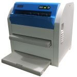
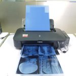
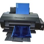
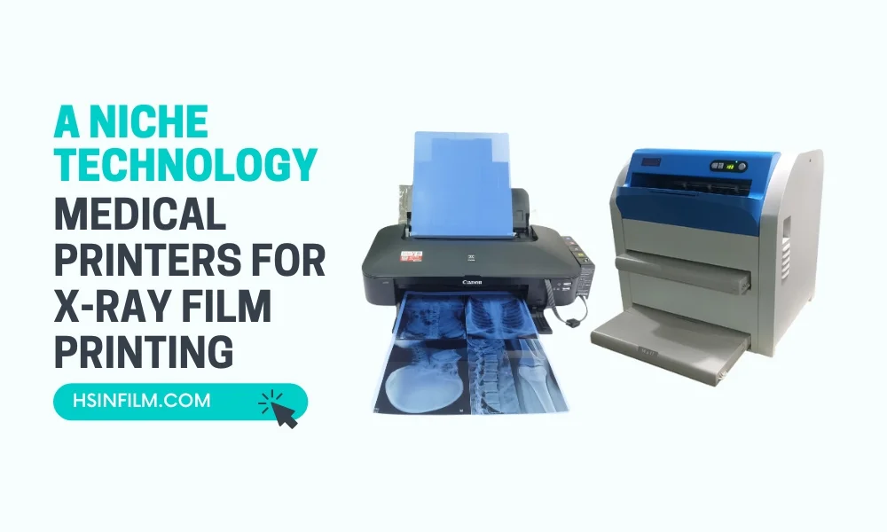
Use Our Best Equipment for Accurate Imaging
HSIN Film is a game-changer in medical imaging, offering precision with their sharp medical dry film, efficiency through quick thermal film processing, and high-quality output with laser film. Their inkjet printers provide versatility, and thermal printers prioritize speed without sacrificing quality. Collectively, HSIN Film ensures accurate and effective diagnostic imaging, enhancing patient care.