Elbow injuries and conditions demand meticulous examination, and the Anteroposterior (AP) Elbow X-ray is a cornerstone in the radiologist’s toolkit. In this comprehensive guide, we’ll delve into the intricacies of interpreting AP Elbow X-rays, providing a detailed handbook for radiologists and medical enthusiasts alike.
Table of Contents
Understanding the Significance of AP Elbow X-rays
Anatomy Spotlight
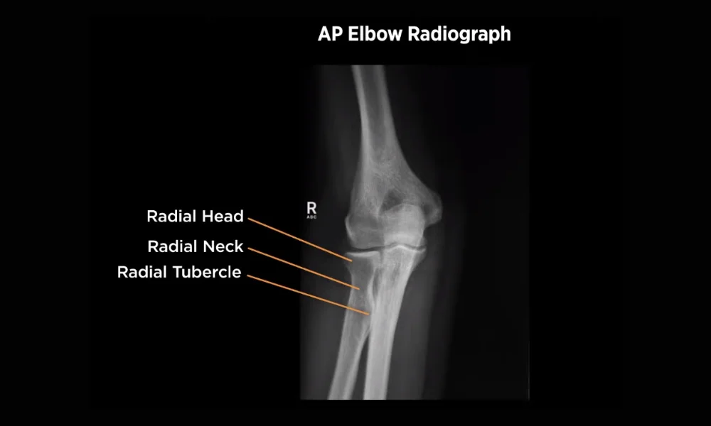
The AP Elbow X-ray captures a frontal view, offering a comprehensive look at the elbow’s intricate anatomy. This includes the humerus, ulna, radius, and associated joints, crucial for detecting fractures, dislocations, and degenerative changes.
Diagnostic Versatility
From traumatic injuries to chronic conditions like arthritis, the AP Elbow X-ray is versatile, serving as a primary diagnostic tool for various elbow-related issues.
Also read: Taking X-ray of Perfect Teeth
Navigating the AP Elbow X-ray: Step-by-Step Analysis
1. Patient Connection
Before delving into the technicalities, connecting with the patient is paramount. Ensuring comfort, explaining the procedure, and addressing concerns create an environment conducive to accurate imaging.
1. Patient Positioning
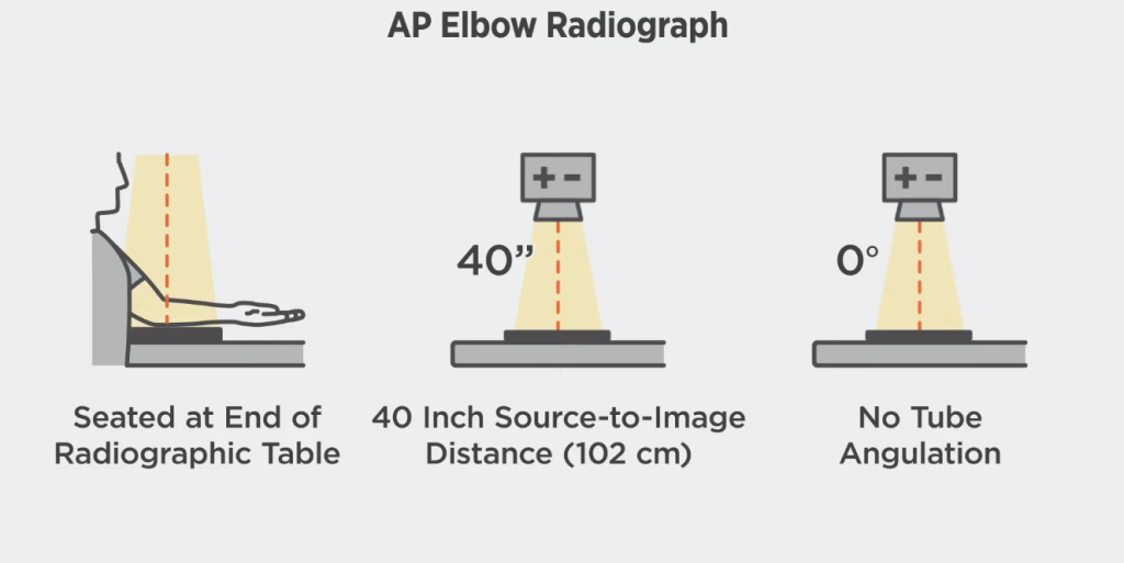
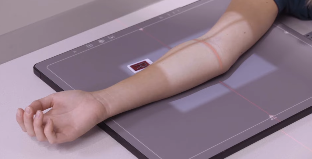
Ensure the patient’s arm is fully extended with the palm facing down. The elbow should be at a right angle, providing an unobstructed view for optimal analysis.
2. Joint Alignment Assessment
Evaluate joint alignment by focusing on the articulation of the humerus, ulna, and radius. Deviations may indicate dislocations or fractures.
3. Bony Structures Examination
Thoroughly inspect bony structures for fractures, paying close attention to the distal humerus, radial head, and olecranon process. Assessing cortical outlines aids in identifying abnormalities.
4. Soft Tissue Evaluation
While primarily focusing on bones, be attentive to soft tissues. Swelling, joint effusion, or calcifications can offer additional diagnostic insights.
Strategies for Accurate Diagnosis Using AP Elbow X-rays
Accurate diagnosis is the cornerstone of effective medical intervention, and when it comes to elbow conditions, Anteroposterior (AP) X-rays play a pivotal role. In this guide, we’ll delve into strategic approaches for radiologists and healthcare professionals to ensure precision in interpreting AP Elbow X-rays, ultimately leading to comprehensive and effective diagnoses.
1. Meticulous Patient Positioning: The Foundation of Precision
- Importance of Alignment: Ensure the patient’s arm is fully extended with the palm facing down, and the elbow forms a right angle. This foundational step guarantees unobstructed views for accurate analysis.
- Optimizing Posture: Attention to detail in patient positioning eliminates potential distortions, providing a clear canvas for radiological interpretation.
2. Systematic Joint Alignment Assessment: Deciphering Symmetry
- Focus on Humerus, Ulna, and Radius: A meticulous evaluation of joint alignment is crucial. Deviations in the articulation of the humerus, ulna, and radius can indicate dislocations or fractures.
- Identifying Discrepancies: Discrepancies in alignment may reveal subtle injuries that could be overlooked without careful scrutiny.
3. Thorough Examination of Bony Structures: Beyond Fracture Detection
- Targeting Key Areas: Thoroughly inspect bony structures, with special attention to potential fracture sites like the distal humerus, radial head, and olecranon process.
- Cortical Outlines for Abnormalities: Assessing cortical outlines aids in identifying abnormalities, providing a more nuanced understanding of the injury’s nature.
4. Soft Tissue Evaluation: Balancing the Skeletal Narrative
- Swelling and Joint Effusion: While bones take center stage, don’t overlook soft tissues. Swelling or joint effusion may offer additional diagnostic clues, contributing to a more comprehensive diagnosis.
- Calcifications and Anomalies: Soft tissue anomalies, such as calcifications, can be subtle indicators of underlying issues, enriching the diagnostic landscape.
5. Pattern Recognition for Common Findings: Interpreting the Story
- Radial Head Fractures: Recognize subtle fractures around the radial head, often associated with specific mechanisms of injury. Understanding fracture patterns aids in accurate diagnosis.
- Supracondylar Fractures: Detecting supracondylar fractures, particularly in pediatric cases, requires a keen eye for subtle deformities and deviations.
6. Advanced Techniques: Elevating Diagnostic Precision
- Stress Views for Ligamentous Assessment: Stress views add dynamics to static images, aiding in the evaluation of ligamentous stability. This advanced technique unveils hidden instabilities not apparent in standard views.
- Contralateral Comparison: Comparing the affected elbow with the contralateral side enhances diagnostic acuity, revealing subtle discrepancies that might escape individual scrutiny.
7. Radiologist’s Expertise: An Art of Interpretation
- Continuous Education: Radiologists must stay updated on the latest advancements and nuances in elbow imaging. Continuous education ensures they remain at the forefront of diagnostic excellence.
- Collaborative Dialogue: Engage in collaborative discussions with orthopedic specialists and clinicians. A synthesis of radiological expertise and clinical insights elevates the accuracy of diagnoses.
8. Leveraging Technology: From Digitalization to AI Assistance
- Digital Radiography for Efficiency: Embrace digital radiography for instant results, reducing waiting times for patients and facilitating quicker diagnoses.
- AI-Assisted Imaging: Explore the integration of artificial intelligence for more nuanced and precise diagnostics. AI can assist in identifying subtle abnormalities and enhancing diagnostic accuracy.
9. Patient-Centric Approach: Enhancing Comfort and Cooperation
- Communication is Key: Clear communication with patients fosters a cooperative environment. Explaining the importance of the procedure and addressing concerns enhances patient cooperation.
- Educational Engagement: Educate patients about the diagnostic process. An informed patient is more likely to cooperate, contributing to clearer images and more accurate assessments.
10. Continuous Improvement: Reflecting on Practices for Enhanced Precision
- Regular Audits and Reviews: Periodic audits of imaging practices and case reviews contribute to continuous improvement. Reflecting on challenging cases enhances the radiologist’s diagnostic acumen.
- Feedback Mechanisms: Establish feedback mechanisms within radiology teams. A collaborative approach to learning from experiences fosters a culture of continuous improvement.
In conclusion, achieving accurate diagnoses using AP Elbow X-rays involves a combination of technical expertise, advanced techniques, and a commitment to continuous improvement. By implementing these strategies, radiologists can enhance their precision, providing patients with thorough and effective diagnostic insights for optimal healthcare outcomes.
Decoding Common Findings in AP Elbow X-rays
1. Radial Head Fractures
Identify subtle fractures around the radial head, often associated with falls on an outstretched hand. Assess displacement and angulation for appropriate management.
2. Supracondylar Fractures
Examine the distal humerus for supracondylar fractures, prevalent in pediatric cases. Recognition is crucial for timely intervention and preventing complications.
3. Olecranon Fractures
Carefully scrutinize the olecranon process for fractures, considering their impact on elbow stability and function. Surgical intervention may be necessary in complex cases.
4. Ligamentous Landscapes
In stress views and comparisons, ligamentous integrity takes center stage. The radiologist traverses the ligamentous landscapes, looking for signs of instability and subtle injuries.
Advanced Techniques and Technologies
1. Stress Views
Consider stress views for subtle ligamentous injuries. Stressing the joint under controlled conditions can unveil instability not apparent in standard views.
2. Comparison with the Contralateral Side
For subtle changes, compare the affected elbow with the contralateral side. Discrepancies in joint spaces or alignment may be more apparent, aiding in diagnosis.
The Role of Radiologist Expertise
1. Pattern Recognition
Years of experience contribute to pattern recognition. Radiologists adept in deciphering AP Elbow X-rays can swiftly identify abnormalities, streamlining the diagnostic process.
2. Collaborative Analysis
Collaborate with orthopedic specialists for a comprehensive assessment. Combining radiological expertise with clinical insights ensures accurate diagnoses and tailored treatment plans.
Conclusion: Mastering AP Elbow X-ray Interpretation
In conclusion, mastering the interpretation of AP Elbow X-rays requires a combination of anatomical knowledge, meticulous analysis, and collaboration with fellow healthcare professionals. Radiologists, armed with this handbook, can enhance their proficiency in deciphering complex elbow conditions, ultimately contributing to improved patient outcomes.
Whether you’re a seasoned radiologist or a medical enthusiast eager to understand the intricacies of diagnostic imaging, decoding AP Elbow X-rays is a rewarding journey into the fascinating realm of musculoskeletal radiology.
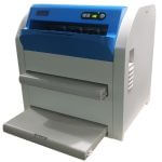
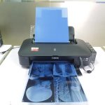
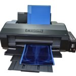
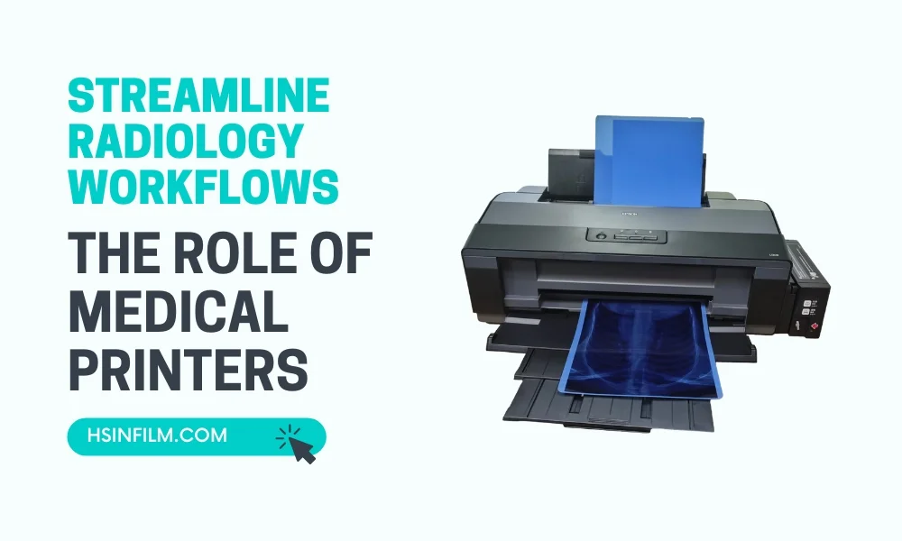
Use HSIN Film’s Products for the Best Accurate Diagnosis
Upgrade your medical imaging from blurry to crystal-clear precision with our Medical Dry Film. Capture temperature-sensitive details with the warm touch of our Medical Thermal Film. Achieve ninja-sharp clarity in your medical images with the precision of our Medical Laser Film. Our Inkjet Printer transforms your health into art, the Thermal Printer crafts every detail delicately, and the Laser Printer orchestrates a symphony of medical imagery. Enjoy top-notch medical imaging at a negotiable price, bringing simplicity, versatility, and user-friendly tech to everyone. Elevate your health; choose not just technology but a commitment to your well-being.