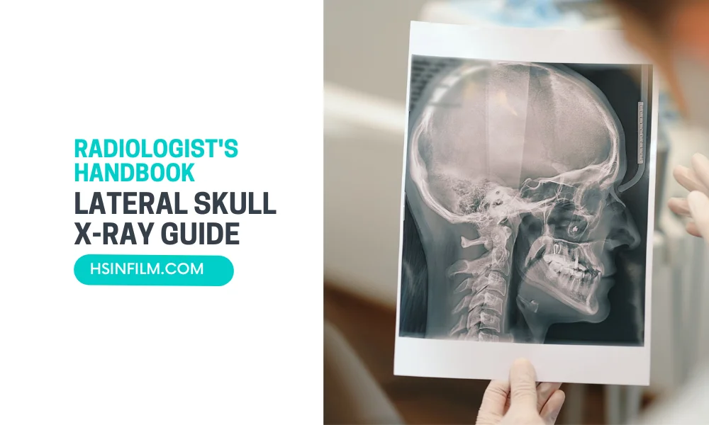
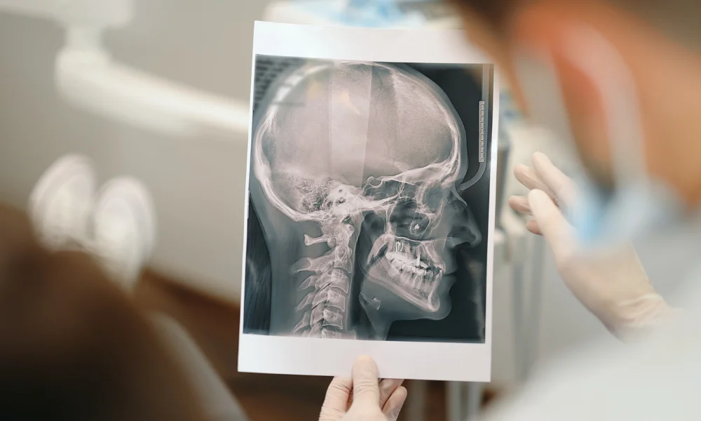
The lateral skull X-ray is a vital diagnostic tool for assessing various conditions affecting the head and cranial structures. In this comprehensive guide, we’ll walk through the intricacies of interpreting lateral skull X-rays, providing radiologists with a handbook for accurate and insightful diagnostics.
Table of Contents
Anatomy of the Skull
The human skull is a complex bony structure that houses and protects the brain, supports sensory organs, and provides the framework for the head and face. It consists of 22 bones divided into two main groups: the cranial bones and the facial bones.
Cranial Bones (8 bones)
These bones form the protective structure around the brain, known as the cranium.
- Frontal Bone: Forms the forehead and the upper part of the eye sockets (orbits).
- Parietal Bones (2): Located on the sides and roof of the cranium.
- Temporal Bones (2): Found below the parietal bones, house structures like the ears and mastoid process.
- Occipital Bone: Forms the back and base of the skull, featuring the foramen magnum (the opening through which the spinal cord passes).
- Sphenoid Bone: A butterfly-shaped bone at the base of the skull, crucial in supporting the brain and connecting the skull with facial bones.
- Ethmoid Bone: Located between the eyes, contributing to the nasal cavity and the orbits.
Facial Bones (14 bones)
These bones give shape to the face and provide cavities for the sensory organs.
- Nasal Bones (2): Form the bridge of the nose.
- Maxillae (2): Upper jawbones that contain the upper teeth and contribute to the orbits.
- Zygomatic Bones (2): Also known as the cheekbones, they connect with the temporal bones.
- Mandible: The lower jawbone, the only movable bone in the skull.
- Lacrimal Bones (2): Small bones forming part of the eye sockets.
- Palatine Bones (2): Form part of the hard palate.
- Inferior Nasal Conchae (2): Located inside the nasal cavity.
- Vomer: Forms part of the nasal septum.
Structures Visualized on a Lateral Skull X-ray
A lateral skull X-ray is an imaging technique that captures a side view of the skull. It is commonly used to assess fractures, tumors, infections, and other abnormalities. The key anatomical structures visible on a lateral skull X-ray include:
- Frontal Bone: Visible at the front of the image, forming the forehead.
- Parietal Bone: Seen on the upper part of the skull, creating the side wall of the cranium.
- Temporal Bone: Clearly visualized below the parietal bone, including the mastoid process.
- Occipital Bone: Found at the back of the skull, extending towards the neck.
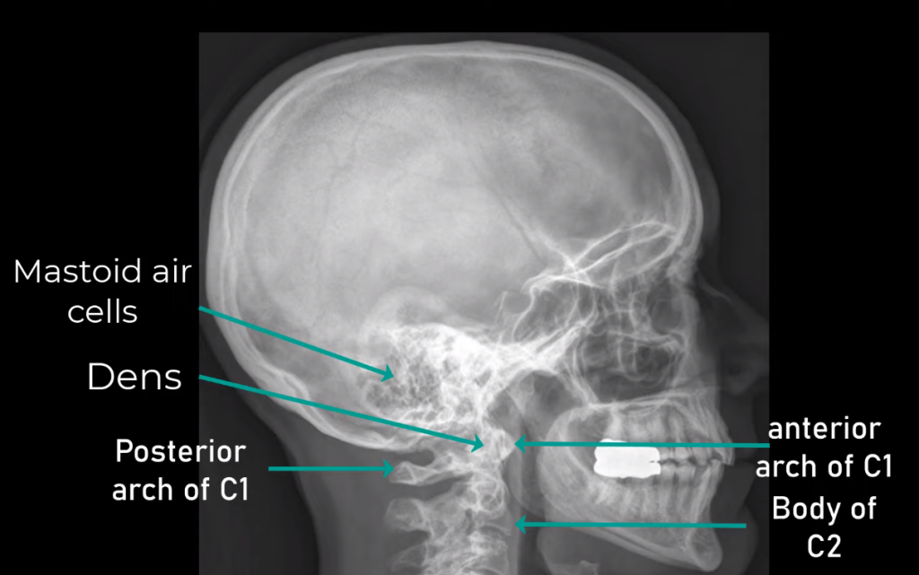
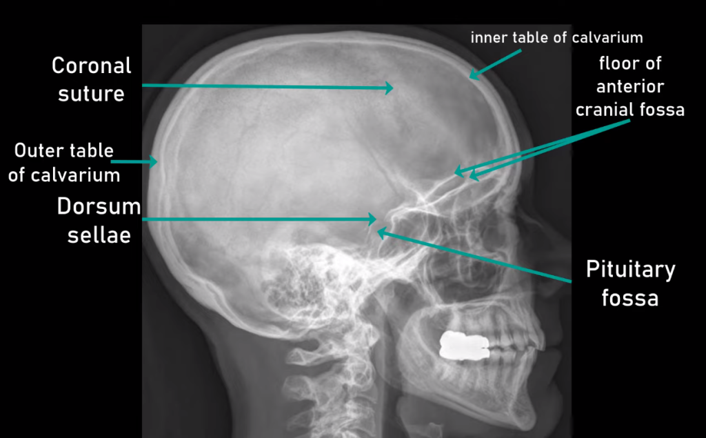
- Sella Turcica: A depression in the sphenoid bone that houses the pituitary gland. This is a critical landmark for assessing intracranial pathology.
- Zygomatic Arch: The cheekbone, forming part of the lateral wall of the orbit.
- Maxilla: The upper jaw, including the roots of the upper teeth.
- Mandible: The lower jaw, including the angle of the mandible, which is often examined for fractures.
- Nasal Cavity: The air-filled space behind the nose.
- Sinuses: The frontal and sphenoid sinuses are visible, providing clues to conditions like sinusitis.
- Orbits: The bony cavities that house the eyes.
- External Auditory Canal: The opening of the ear canal within the temporal bone.
- Cervical Spine: The upper part of the cervical vertebrae (C1 and C2) may be partially visualized at the base of the skull, particularly in trauma cases.
- Foramen Magnum: The large opening at the base of the skull, though partially obscured, where the spinal cord connects to the brainstem.
Clinical Applications of a Lateral Skull X-ray
- Fractures: Identifying fractures in the cranial bones, particularly after trauma.
- Infections: Assessing signs of mastoiditis or other bone infections.
- Tumors: Detecting abnormal growths that may affect the bones or soft tissues.
- Sinus Problems: Visualizing air-fluid levels or blockages in the frontal or sphenoid sinuses.
- Hydrocephalus: Evaluating the sella turcica for signs of raised intracranial pressure.
- Developmental Disorders: Assessing abnormal skull shapes in conditions like craniosynostosis.
Importance of a Lateral Skull X-ray
A lateral skull X-ray provides a clear side view of the skull, allowing clinicians to examine the structural integrity and detect abnormalities in various anatomical regions. It is often one of the first imaging tests ordered in trauma cases and continues to be valuable in diagnosing infections, tumors, and congenital anomalies.
Indications of Lateral Skull X-ray
A lateral skull X-ray is a crucial diagnostic tool in various clinical conditions, especially when evaluating the bones of the skull, facial structures, and certain intracranial abnormalities. It is often one of the initial imaging modalities used in the following clinical scenarios:
1. Head Trauma
- Indication: A lateral skull X-ray is commonly ordered when a patient experiences head trauma, especially if there is a suspicion of a skull fracture or other bone-related injuries.
- Information Provided: It helps identify fractures in the cranial bones, such as the frontal, parietal, and occipital bones. The X-ray can also show air or fluid levels inside the cranial cavity, which may suggest intracranial bleeding or cerebrospinal fluid leakage.
- Associated Conditions:
- Linear Skull Fractures: The X-ray helps detect non-displaced fractures, which may increase the risk of intracranial injury.
- Depressed Skull Fractures: Visible indentations in the skull, where broken bone segments press into the brain, may require surgical intervention.
2. Facial Fractures
- Indication: Facial trauma, especially involving the cheekbones (zygomatic arches), nasal bones, or mandible, may necessitate a lateral skull X-ray for assessment.
- Information Provided: The X-ray helps in identifying fractures in the facial bones and determining the severity of the injury. It also assesses the alignment and possible displacement of the bones.
- Associated Conditions:
- Zygomatic Fractures: A lateral view is helpful to assess fractures of the cheekbone and their effect on the eye socket (orbit).
- Nasal Bone Fractures: Common in facial trauma, especially from sports injuries or accidents, the lateral skull X-ray helps in confirming the presence of fractures in the nasal bones.
- Mandibular Fractures: Fractures of the lower jaw, which may affect dental alignment and require surgical correction.
3. Intracranial Mass Lesions
- Indication: In patients with symptoms suggesting an intracranial mass (such as headaches, seizures, or focal neurological deficits), a lateral skull X-ray may be performed as an initial step before advanced imaging like CT or MRI.
- Information Provided: Although X-rays provide limited direct information about soft tissue structures, they can reveal secondary signs of intracranial masses, such as changes in bone structure or increased intracranial pressure.
- Associated Conditions:
- Tumors: A mass effect on the skull bones or expansion of the sella turcica (indicating pituitary tumors) can be visible on a lateral X-ray.
- Hydrocephalus: Enlarged cranial sutures or changes in skull shape due to increased intracranial pressure can suggest conditions like hydrocephalus.
- Meningioma: Some meningiomas can cause hyperostosis (thickening of the skull bone), which may be seen on an X-ray.
4. Sinus Infections and Conditions
- Indication: Chronic sinus infections or suspected sinusitis can be assessed using a lateral skull X-ray, especially if the frontal or sphenoid sinuses are involved.
- Information Provided: Air-fluid levels in the sinuses or sinus opacification can indicate infection or inflammation.
- Associated Conditions:
- Sinusitis: Inflammation or infection of the paranasal sinuses is commonly visible on X-rays as fluid accumulation or mucosal thickening.
- Sinus Tumors: Although rare, tumors of the sinuses can cause bone destruction or mass effect, which can be visualized on a lateral skull X-ray.
5. Developmental Abnormalities
- Indication: Congenital or developmental conditions affecting the shape or growth of the skull may warrant a lateral skull X-ray to assess cranial sutures and bone alignment.
- Information Provided: The X-ray allows the visualization of abnormal suture closure or bone malformations that may indicate conditions like craniosynostosis or other craniofacial disorders.
- Associated Conditions:
- Craniosynostosis: Premature fusion of the cranial sutures, leading to abnormal skull shape and potential developmental delays, can be detected.
- Microcephaly: A smaller-than-normal skull size, associated with developmental disorders, can also be confirmed through lateral skull X-ray.
6. Temporomandibular Joint (TMJ) Disorders
- Indication: Patients with jaw pain, difficulty opening or closing their mouth, or clicking/popping sounds in the jaw may undergo a lateral skull X-ray to assess the TMJ.
- Information Provided: The X-ray shows the bony structure of the TMJ and can help in identifying displacement or degeneration of the joint.
- Associated Conditions:
- TMJ Dysfunction: Changes in the alignment or structure of the joint due to arthritis or trauma can be evaluated.
7. Osteomyelitis and Bone Infections
- Indication: Suspected bone infections, particularly in the skull, can prompt the use of a lateral skull X-ray for evaluation.
- Information Provided: The X-ray helps detect bone destruction or irregularities caused by infection.
- Associated Conditions:
- Osteomyelitis of the Skull: An infection in the cranial bones, often secondary to trauma or sinusitis, may show bone erosion or periosteal reaction on an X-ray.
A lateral skull X-ray provides valuable insights into conditions affecting the cranial and facial bones. It serves as an essential first-line imaging technique in trauma, infection, tumors, developmental abnormalities, and TMJ disorders. While advanced imaging modalities like CT and MRI are often used for more detailed evaluations, the lateral skull X-ray remains a simple, accessible tool for detecting structural abnormalities and guiding further treatment decisions.
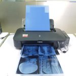
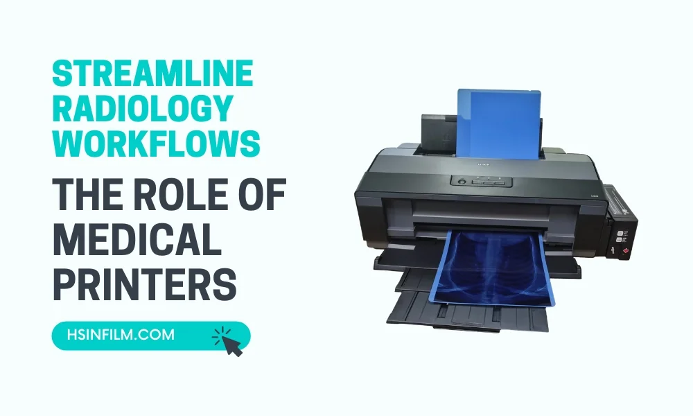
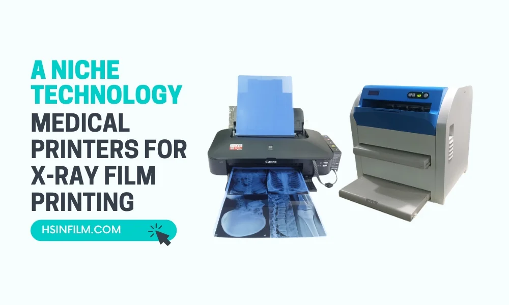
Fracture lines and subtle findings are easiest to catch on high-contrast film. HSIN’s Medical Laser Film and Medical Thermal Film are DICOM-compatible and built for consistent diagnostic quality — with free setup support included.
Get a Free QuoteAlso read: Understanding the Significance of Lateral Skull X-rays
Implications of a Lateral Skull X-ray
A lateral skull X-ray provides important diagnostic information by visualizing the bones and some soft tissues of the head and face. Though modern imaging techniques like CT and MRI offer more detailed views, lateral skull X-rays remain valuable for certain conditions due to their ability to quickly and effectively identify specific abnormalities. Here are the key diagnostic implications:
1. Fractures and Bone Abnormalities
- Linear Skull Fractures: Lateral skull X-rays are useful for detecting linear skull fractures that might be less obvious on a CT scan, especially in trauma cases. They can reveal fractures along the parietal, frontal, and occipital bones.
- Depressed Fractures: In some cases, lateral X-rays provide clear images of depressed fractures where segments of the skull are pressed inward, possibly impacting the brain tissue. These fractures may be more subtle or missed on other imaging modalities.
- Cranial Sutures: This imaging technique also provides a clear view of cranial sutures, helping diagnose conditions like craniosynostosis, where premature closure of sutures can lead to abnormal skull growth.
2. Tumors and Masses
- Bone Erosion from Tumors: In cases where intracranial or skull-based tumors affect the bone, a lateral skull X-ray can show bone erosion or hyperostosis (thickening of bone), particularly in conditions like meningioma. While MRI is more commonly used to detect tumors, X-rays are still useful in visualizing how a tumor may be affecting the skull structure.
- Pituitary Tumors: Lateral skull X-rays can reveal enlargement or erosion of the sella turcica, a bony structure where the pituitary gland sits. This can be a sign of pituitary adenomas or other pituitary tumors, which may not be easily visible in early stages with other imaging techniques.
3. Air or Fluid Levels
- Sinus Disease: Lateral skull X-rays are helpful in assessing the paranasal sinuses for air-fluid levels that indicate conditions like sinusitis. These air-fluid levels may not be immediately apparent in more detailed imaging, where the focus is on soft tissues rather than gas and fluid accumulations.
- Intracranial Hemorrhage: In trauma cases, air or fluid in spaces within the skull (e.g., epidural or subdural space) may be visible on a lateral X-ray, providing an early clue to the presence of intracranial hemorrhage or CSF leakage before further testing with CT scans.
4. Developmental Anomalies
- Craniosynostosis: This condition involves the premature fusion of skull sutures in infants and children, leading to abnormal head shapes and developmental concerns. A lateral skull X-ray can detect abnormal suture closure, allowing early diagnosis and intervention, sometimes more clearly than an ultrasound.
- Microcephaly: The lateral skull X-ray can help assess skull size in relation to age norms, offering a straightforward and quick means of diagnosing microcephaly, where the skull and brain are smaller than expected.
5. Temporomandibular Joint (TMJ) Disorders
- Bony Changes: A lateral skull X-ray can visualize the TMJ and reveal bony changes in patients with TMJ disorders. It helps identify degenerative changes in the joint structure, dislocations, or trauma-related damage. Though an MRI provides better detail for soft tissues like the articular disc, the X-ray is useful for assessing the bones of the jaw and skull base.
6. Dental and Maxillofacial Applications
- Facial Fractures: Lateral skull X-rays play a crucial role in diagnosing fractures of the zygomatic arch, mandible, and maxilla. These fractures may not always be visible on standard dental X-rays but can be clearly seen on a lateral skull view.
- Dental Malocclusions: Lateral skull X-rays help orthodontists and dentists assess malocclusions (misaligned teeth) by providing detailed views of the alveolar ridge and the relationship between the upper and lower jaws.
7. Intracranial Pressure and Hydrocephalus
- Hydrocephalus: A lateral skull X-ray can indicate increased intracranial pressure due to hydrocephalus (accumulation of fluid in the brain). The imaging can show thinning of the cranial bones, enlargement of the sutures, or even erosion of the inner skull layers, providing evidence of underlying pressure buildup that might not be immediately apparent in non-radiographic tests.
8. Vascular Abnormalities
- Calcifications: Some vascular abnormalities, such as calcified carotid arteries or pineal gland calcifications, can be identified on a lateral skull X-ray. These calcifications may not be as visible on soft-tissue-focused imaging like MRI, making X-rays valuable for identifying mineralized structures in the brain.
Advantages Over Other Imaging Modalities
- Speed and Accessibility: Lateral skull X-rays are quick to perform and widely available, making them an essential first-line diagnostic tool, especially in emergency or trauma settings where speed is crucial.
- Bone Imaging: While CT scans and MRIs provide detailed information on soft tissues, X-rays excel at capturing fine details of bone structure. Fractures, dislocations, and bone lesions are more easily visible on X-rays than on MRI, which primarily targets soft tissues.
- Cost-Effectiveness: X-rays are much less expensive than CT or MRI scans, making them a more accessible option for initial imaging.
Limitations Compared to Other Imaging Modalities
- Limited Soft Tissue Visualization: Unlike MRI, which provides excellent contrast for soft tissues like brain matter, nerves, and blood vessels, lateral skull X-rays have limited use in diagnosing soft tissue abnormalities.
- Two-Dimensional Imaging: X-rays provide a 2D representation of structures, which may sometimes obscure deeper abnormalities. CT and MRI scans offer 3D reconstructions and cross-sectional views, allowing more comprehensive assessments.
Lateral skull X-rays continue to serve an essential role in diagnosing various conditions affecting the skull, face, and underlying structures. While advanced imaging modalities like CT and MRI offer more detailed soft tissue imaging, X-rays are highly valuable for detecting bone abnormalities, fractures, sinus issues, and cranial deformities. Their affordability, speed, and accessibility ensure their continued use in both emergency and routine clinical settings.
Navigating the Lateral Skull X-ray: Step-by-Step Analysis
Patient Positioning for Precision
- Achieving Lateral View: Ensure the patient is in lateral position with the head in true lateral alignment. Proper alignment guarantees optimal imaging of cranial structures.
- Minimizing Overlapping Structures: Adjust the patient’s position to minimize overlapping structures, enhancing clarity in the lateral view.
Evaluating Cranial Structures
- Cranial Bone Assessment: Thoroughly examine the cranial bones for fractures, irregularities, or signs of trauma.
- Sinus Inspection: Focus on sinus areas for signs of inflammation, opacification, or air-fluid levels.
Analyzing Facial Structures
- Facial Bone Integrity: Assess the integrity of facial bones, particularly around the orbits and nasal structures.
- Soft Tissue Examination: Soft tissue analysis reveals potential anomalies, providing a holistic understanding of cranial health.
Diagnostic Value in Trauma Cases
- Identify Fractures: Crucial in identifying fractures and assessing the severity of head injuries in trauma cases.
- Assists: Guides treatment decisions and potential surgical interventions.
Contributions to Neurological Diagnoses
- Neurological Contribution: Beyond bone-related issues, supports neurological diagnoses by identifying abnormalities in the brain’s shape, size, or position.
- Special Cases: Especially useful in assessing conditions like hydrocephalus.
Limitations and Complementary Imaging
Has limitations in visualizing soft tissues; complementary imaging like CT scans or MRI may be recommended for a comprehensive evaluation.
Radiation Safety Measures
Emphasizes the importance of radiation safety, incorporating lead shielding and optimizing exposure settings to minimize radiation exposure.
Interpreting Radiographic Findings
- Requirements: Requires expertise in recognizing normal anatomy and identifying abnormalities.
- Role of Radiologists: Radiologists play a crucial role in providing accurate and detailed reports for informed clinical decision-making.
Navigating the landscape of lateral skull X-rays in radiology demands technical precision and interpretative skill, making them invaluable tools for diagnosing various conditions and contributing to comprehensive patient care.
Deciphering Common Findings in Lateral Skull X-rays
1. Normal Skull Anatomy
- Recognizing Symmetry: Normal Cranial Features showcase a balanced appearance of cranial bones.
- Clear Delineation: The skull base and vault should be distinctly outlined.
- Expected Contours: Familiarize with the typical contours of facial bones.
2. Fractures and Trauma Indicators
- Cracks and Signs of Impact: Identify linear or depressed fractures and signs of impact.
- Disrupted Bone Continuity: Assess any break in the normal bone structure.
- Abnormal Alignment: Recognize bones not aligned as expected.
3. Sinus Abnormalities
- Beyond the Surface: Spot Sinus Congestion, Inflammation, and Blockages.
- Assessment of Paranasal Sinuses: Detect congestion, inflammation, or blockages.
- Contribution to Diagnosis: Aid in diagnosing conditions like sinusitis.
4. Developmental Anomalies
- Unusual Growth Patterns: Recognize anomalies or irregular growth patterns.
- Identification of Asymmetries: Spot any asymmetries in cranial development.
- Vital for Early Intervention: Early detection is crucial for effective management.
5. Hydrocephalus Signs
- Fluid on the Brain: Detect indications of hydrocephalus.
- Abnormal CSF Accumulation: Recognize abnormal cerebrospinal fluid accumulation.
- Early Diagnosis: Contribute to the early diagnosis of hydrocephalus.
6. Tumors and Abnormal Masses
- Beyond the Bone: Recognize Intracranial Tumors and Abnormal Masses.
- Contribution to Detection: Aid in the early detection of intracranial tumors.
- Initial Insights: Provide initial insights, prompting further investigation.
7. Vascular Abnormalities
- Blood Flow Anomalies: Identify irregularities in blood flow patterns.
- Vessel Calcifications: Recognize calcifications in blood vessels.
- Prompting Further Evaluation: Indicate potential circulatory issues, prompting further evaluation.
8. Skull Base Abnormalities
- Foundation Issues: Assess abnormalities in the skull base.
- Understanding Anatomical Variations: Recognize anatomical variations or abnormalities.
- Guiding Clinicians: Help clinicians comprehend foundational aspects of cranial structure.
9. Soft Tissue Abnormalities
- Not Just Bones: Recognize Issues in Soft Tissues Alongside Bones.
- Primary Focus on Bones: While bones are primary, notice any soft tissue swelling or masses.
- Indication of Underlying Issues: Soft tissue abnormalities may indicate underlying issues requiring comprehensive assessment.
10. Artifacts and Technical Considerations
- Navigating the Noise: Distinguish True Findings from Artifacts.
- Understanding Potential Artifacts: Be aware of potential artifacts in lateral skull X-rays.
- Ensuring Accurate Interpretation: Distinguish true findings from technical irregularities, ensuring accurate interpretation and avoiding misdiagnosis.
Advanced Strategies for Analyzing Lateral Skull X-rays
Analyzing lateral skull X-rays requires a nuanced approach, especially when delving into complex cases. Radiologists, equipped with advanced strategies, can extract richer diagnostic insights from this imaging modality. Let’s explore these advanced techniques for a more thorough interpretation.
1. Multi-Planar Analysis: Beyond Lateral Views
- Sagittal and Coronal Reconstructions: Incorporate sagittal and coronal reconstructions alongside the lateral view for a comprehensive three-dimensional analysis.
- Enhanced Anatomical Context: Multi-planar analysis provides enhanced anatomical context, aiding in the identification of subtle abnormalities.
2. Dynamic Imaging Techniques
- Flexion and Extension Views: Implement dynamic imaging with flexion and extension views, especially in trauma cases. This reveals instability and abnormal movements not apparent in static lateral images.
- Stress Testing for Ligaments: Stress testing helps evaluate ligamentous stability, crucial in cases where ligament injuries might contribute to clinical symptoms.
3. Contrast-Enhanced Imaging
- Intravenous Contrast for Vascular Assessment: In selected cases, consider contrast-enhanced imaging for vascular assessment, particularly when investigating vascular anomalies or pathology.
- Identification of Lesions: Contrast-enhanced studies enhance lesion identification, making them more conspicuous against surrounding tissues.
4. Quantitative Analysis Tools
- Densitometry for Bone Density Measurements: Utilize densitometry tools for precise bone density measurements. This is particularly valuable in assessing conditions affecting bone mineralization.
- Quantitative Assessment of Soft Tissues: Employ quantitative tools for the assessment of soft tissue densities, aiding in the identification of subtle abnormalities.
5. Advanced Imaging Modalities Integration
- MRI and CT Fusion: Integrate MRI or CT data with lateral skull X-rays for a more comprehensive evaluation, especially in cases requiring detailed soft tissue assessment.
- PET-CT Correlation: In cases with suspected metabolic activity, correlate PET-CT findings with lateral skull X-rays for a more holistic understanding of pathology.
6. Computer-Aided Detection (CAD) Systems
- Automated Fracture Detection: Implement CAD systems for automated fracture detection, allowing for faster identification of fractures, especially in trauma scenarios.
- Lesion Recognition: CAD systems aid in lesion recognition, acting as a valuable second pair of eyes to ensure no subtle abnormalities go unnoticed.
7. Texture Analysis for Soft Tissues
- Soft Tissue Characterization: Apply texture analysis algorithms for soft tissue characterization. This enhances the differentiation of various soft tissue components, contributing to more accurate diagnoses.
8. Quantitative Metrics for Symmetry Analysis
- Symmetry Metrics Calculation: Employ quantitative metrics to calculate symmetry indices, aiding in the identification of asymmetries or subtle deviations from the norm.
- Objective Comparison: Quantitative analysis provides an objective basis for comparing structures, minimizing subjectivity in interpretation.
9. Educational Platforms for Continuous Learning
- Virtual Case Libraries: Engage with virtual case libraries featuring diverse lateral skull X-ray cases. This continuous exposure enhances diagnostic acumen.
- Interactive Learning Modules: Interactive learning modules facilitate ongoing education, allowing radiologists to stay abreast of evolving trends and techniques.
Implementing these advanced strategies transforms lateral skull X-ray analysis into a sophisticated diagnostic tool. Radiologists, armed with these techniques, can navigate through intricate cases with heightened precision, ensuring optimal patient care and diagnostic accuracy.
Techniques for Accurate Interpretation of Lateral Skull X-rays

1. Alignment and Symmetry Check
- Central Line Alignment: Ensure the central line aligns with the midline of the face.
- Bilateral Symmetry: Confirm symmetry in facial bones, indicating normal development.
2. Bone Density Assessment
- Homogeneous Density: Observe for consistent bone density throughout the skull.
- Identification of Abnormalities: Identify areas of increased or decreased density, indicating potential issues.
3. Soft Tissue Analysis
- Tissue Contour Evaluation: Assess the contour of soft tissues, especially in the neck and facial regions.
- Recognition of Swelling or Masses: Identify any soft tissue abnormalities, such as swelling or masses.
4. Systematic Bone Evaluation
- Sequential Bone Assessment: Systematically evaluate each bone from the base to the vault.
- Identification of Fractures or Anomalies: Detect any fractures, structural anomalies, or irregularities.
5. Recognition of Normal Landmarks
- Key Landmark Identification: Recognize and identify normal anatomical landmarks, ensuring proper orientation.
- Guidance for Interpretation: Landmarks provide guidance, aiding in the interpretation of surrounding structures.
6. Review of Nasal Structures
- Nasal Septum Assessment: Examine the nasal septum for alignment and potential deviations.
- Identification of Sinus Abnormalities: Assess paranasal sinuses for any signs of inflammation or abnormalities.
7. Evaluation of Cranial Base
- Stability Check: Confirm stability and alignment of the cranial base.
- Detection of Abnormalities: Identify any abnormalities in the bones forming the cranial base.
8. Assessment of Skull Vault
- Recognition of Abnormalities: Examine the skull vault for signs of deformities, lesions, or irregularities.
- Understanding Developmental Variations: Differentiate between normal developmental variations and potential issues.
9. Recognition of Vascular Patterns
- Vascular Pathway Identification: Recognize normal vascular patterns within the skull.
- Detection of Anomalies: Identify any irregularities or calcifications in blood vessels.
Potential Risks and Benefits of a Lateral Skull X-ray
A lateral skull X-ray is a common diagnostic tool used to visualize the bones and some soft tissues of the head. While it remains an essential part of medical imaging, like any procedure, it comes with its own set of risks and benefits. Here’s a detailed exploration of both:
Benefits of a Lateral Skull X-Ray
Quick and Accessible Diagnosis
- Speed: Lateral skull X-rays can be performed quickly, often taking only a few minutes. This makes them an ideal first-line imaging tool, especially in emergency or trauma situations.
- Accessibility: Unlike CT or MRI machines, which are more complex and less widely available, X-ray equipment is commonly found in most hospitals and clinics, making it a readily accessible imaging option.
Effective for Detecting Bone Fractures and Abnormalities
- Fractures: Lateral skull X-rays are especially useful for detecting linear skull fractures, depressed fractures, and cranial bone lesions. These are conditions where speed and simplicity of diagnosis are critical.
- Developmental Conditions: It can help detect developmental anomalies like craniosynostosis (premature closure of cranial sutures) and microcephaly (abnormally small head size), which may be easier to identify on X-rays than on other imaging modalities.
Cost-Effective
- Affordability: Compared to CT or MRI scans, X-rays are significantly more cost-effective. This makes them a preferred imaging choice in situations where expensive imaging methods are not necessary or are unavailable.
Low Radiation Exposure Compared to Other Imaging Modalities
- While X-rays do involve exposure to ionizing radiation, the amount is considerably lower compared to CT scans, making it a safer option for patients when multiple images or repeated scans are required.
Visualization of Specific Conditions
- Bone Erosion: In conditions like tumors or bone infections (e.g., osteomyelitis), lateral skull X-rays can reveal areas of bone erosion, a key indicator of disease.
- Sinus Disease: It can help identify air-fluid levels in the paranasal sinuses, indicating infections or sinusitis.
- Vascular Abnormalities: Calcifications of structures like the carotid artery or pineal gland can sometimes be visualized on a lateral skull X-ray.
Minimal Patient Preparation
- Simplicity: Unlike other imaging tests that may require the patient to fast, be sedated, or inject contrast material, lateral skull X-rays generally require little to no preparation.
Risks of a Lateral Skull X-Ray
Radiation Exposure
- Ionizing Radiation: Like all X-rays, lateral skull X-rays expose patients to ionizing radiation, which can increase the risk of cancer over time, particularly with repeated exposure. While the dose from a lateral skull X-ray is relatively low, it is still a risk that must be considered.
- Cumulative Effects: For individuals who require multiple X-rays or other radiation-based imaging, the cumulative dose could pose a greater risk, especially in more vulnerable populations like children and pregnant women.
Limited Information on Soft Tissues
- Soft Tissue Inadequacy: Lateral skull X-rays are effective for bones but offer limited detail on soft tissues such as the brain, blood vessels, or nerves. Conditions like intracranial bleeding, tumors, or vascular abnormalities are more difficult to detect compared to advanced imaging techniques like CT or MRI.
- Missed Diagnoses: Certain intracranial conditions, such as stroke, tumor growth, or cerebral hemorrhage, may go undetected on an X-ray but would be visible on more advanced imaging like a CT scan or MRI.
Two-Dimensional Imaging
- Lack of Depth: Lateral skull X-rays provide a 2D view of the head and skull, making it difficult to assess abnormalities that may be hidden behind other structures. This limitation sometimes necessitates follow-up imaging with CT or MRI for a more comprehensive evaluation.
Potential for Diagnostic Errors
- Positioning Errors: If the patient is not positioned correctly or moves during the procedure, the X-ray may result in poor-quality images, which can lead to misinterpretation or misdiagnosis.
- Artifacts: Any metallic objects, such as earrings or dental fillings, can create artifacts that interfere with the image and potentially obscure important diagnostic information.
Not Suitable for All Conditions
- Other Modalities Preferred: For conditions involving soft tissue, vascular disorders, or brain injuries, lateral skull X-rays are not as informative as a CT scan or MRI, limiting their utility for certain medical conditions.
Pregnancy Concerns
- Fetal Exposure: Pregnant women are generally advised to avoid unnecessary X-rays to minimize the risk of radiation exposure to the developing fetus, even though the radiation dose is low. Shielding may be required to reduce exposure in certain cases.
Risk-Benefit Analysis
- For Trauma: In emergency settings where speed is crucial, the benefits of quickly identifying fractures or other bone abnormalities often outweigh the minimal risks of radiation exposure. However, follow-up imaging with a CT scan or MRI may still be needed.
- For Developmental Conditions: The lateral skull X-ray is a valuable, cost-effective tool for identifying abnormalities like premature suture closure (craniosynostosis) or bone growth problems, where the benefits clearly outweigh the risks.
- Routine Use: For conditions requiring frequent monitoring, such as tumor surveillance, the cumulative radiation exposure must be carefully considered, and alternative imaging methods like MRI may be preferable.
A lateral skull X-ray is a valuable, accessible, and cost-effective tool in medical imaging, particularly for diagnosing bone fractures, developmental conditions, and sinus diseases. The procedure carries minimal radiation exposure compared to other imaging techniques like CT scans. However, its limitations in soft tissue imaging, risk of radiation exposure, and potential for diagnostic errors should be weighed carefully. When used appropriately, its benefits far outweigh the risks, particularly in trauma or emergency settings.
Conclusion: Mastering Lateral Skull X-ray Interpretation
In conclusion, mastering on navigating, deciphering common findings, strategies for analyzing, and techniques of interpretation of lateral skull X-rays demands a blend of technical proficiency, anatomical knowledge, and a commitment to continuous learning. This guide serves as a companion for radiologists, providing insights into the nuances of lateral skull X-ray interpretation and contributing to enhanced diagnostic precision.