Root canal treatments are vital for preserving teeth that are severely infected or decayed. One essential tool in diagnosing and planning for root canal therapy is the X-ray. Dental X-rays provide a clear view of the internal structure of the tooth and the surrounding areas, enabling dentists to identify infections, fractures, and other issues. This blog post will explore the root canal X-ray recommendation where X-rays are necessary before, during, and after root canal treatment.
Table of Contents
What is a Root Canal?
Understanding Root Canal Therapy
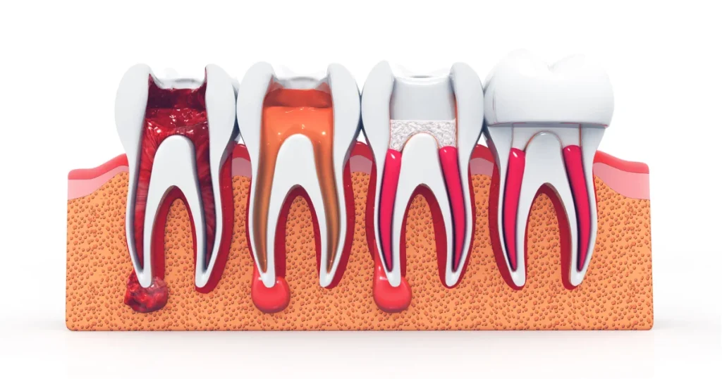
A root canal is a dental procedure aimed at treating the infection or damage to the pulp of a tooth, which is the innermost layer that contains nerves and blood vessels. During this treatment, the dentist removes the infected or damaged pulp, cleans the inside of the tooth, and seals it to prevent further infection.
Root canals are usually recommended when a tooth is severely decayed, injured, or infected, causing pain, swelling, or abscesses. To determine the extent of the infection or damage, dental X-rays are often necessary.
Role of X-rays in Root Canal Therapy
X-rays play an essential role in root canal treatment by providing dentists with a clear view of the affected tooth and surrounding bone. They help in diagnosing the problem, guiding the treatment, and evaluating the success of the procedure. Let’s take a closer look at when and why X-rays are used.
Also read: A Comprehensive Guide Through Root Canal X-rays
5 Root Canal X-ray Recommendations
1. Diagnosing the Need for a Root Canal
One of the most common reasons dentists recommend X-rays is to diagnose whether a root canal is needed. A regular examination may not provide enough information about the condition of the tooth’s pulp or the extent of decay, especially if it is hidden beneath the gum line.
Key Reasons for Diagnostic X-rays:
- Severe Tooth Pain: If a patient is experiencing intense pain, particularly when biting or applying pressure to the tooth, an X-ray can help identify the source of the pain, such as an abscess or deep decay.
- Sensitivity to Hot or Cold: Prolonged sensitivity to temperature can be a sign of pulp damage. An X-ray will show whether the damage extends to the root of the tooth, indicating the need for a root canal.
- Swelling or Abscess: X-rays are also useful in detecting abscesses, which are pockets of pus that form at the root of an infected tooth. These abscesses may not always be visible or felt, but they can be detected through X-rays.
- Hidden Decay: Sometimes, decay can spread beneath a filling or crown, making it difficult to spot during a regular exam. X-rays can reveal decay that’s not visible to the naked eye.
2. Planning the Root Canal Procedure
Once it’s determined that a root canal is needed, additional X-rays are taken to plan the procedure. The dentist needs a detailed image of the tooth’s structure, including the roots and surrounding bone, to ensure that the treatment is effective and minimizes complications.
Reasons for Pre-Treatment X-rays:
- Mapping the Root Canals: Teeth can have more than one root canal, and their shapes can vary significantly. X-rays help in identifying all the canals and their pathways, which is crucial for removing all the infected tissue.
- Checking the Length of the Root: X-rays also show the length of the tooth’s root. This information is vital to ensure that the dentist cleans and seals the entire canal properly.
- Assessing Bone Structure: Before beginning the procedure, the dentist may also need to evaluate the surrounding bone for signs of infection or damage. This helps in determining the severity of the infection and planning the best course of action.
3. Monitoring Progress During the Procedure
During a root canal procedure, dentists may take X-rays at various stages to ensure that the treatment is progressing smoothly. These X-rays provide real-time feedback and help the dentist confirm that the infected tissue has been fully removed and that the root canals have been properly cleaned and shaped.
Reasons for Mid-Treatment X-rays:
- Ensuring Complete Removal of Infection: The dentist may take an X-ray to confirm that all infected or damaged tissue has been removed from the root canals.
- Checking Instrument Placement: X-rays are often used to check the placement of dental instruments within the canals, ensuring that they are properly positioned and not damaging the surrounding tissues.
- Confirming Proper Filling: Once the canals are cleaned, they are filled with a special material. An X-ray may be taken to confirm that the filling material has been placed correctly, without any gaps or overfilling.
4. Post-Treatment Follow-Up
After the root canal procedure is complete, X-rays are taken to ensure that the treatment was successful and that the tooth is healing properly. These post-treatment X-rays are vital for detecting any remaining infection or complications, such as overfilled canals or damage to the surrounding bone.
Reasons for Post-Treatment X-rays:
- Verifying Treatment Success: An X-ray after the procedure helps the dentist confirm that the root canal has been thoroughly cleaned and sealed.
- Checking for Complications: Sometimes, complications can arise after a root canal, such as an infection that wasn’t completely removed or a crack in the tooth. Follow-up X-rays can help detect these issues early on, allowing for prompt treatment.
- Monitoring Healing: The dentist may also use X-rays to monitor the healing of the surrounding bone and tissues, especially if there was an abscess or significant infection before the procedure.
5. Regular Check-Ups After a Root Canal
Even after a successful root canal treatment, regular dental check-ups are essential to ensure that the tooth remains healthy and free of infection. X-rays may be recommended during these check-ups to monitor the condition of the treated tooth and to ensure that no new problems have developed.
Key Reasons for Routine X-rays After a Root Canal:
- Detecting Recurrent Infections: In some cases, a tooth that has undergone root canal treatment may become reinfected. Routine X-rays help in detecting any signs of infection early on, allowing for timely intervention.
- Monitoring Bone Health: X-rays can also help in monitoring the health of the surrounding bone, ensuring that it is healing properly and that no bone loss is occurring.
- Checking for Structural Integrity: Over time, the structural integrity of a tooth that has undergone root canal therapy can weaken. X-rays help in assessing the strength of the tooth and determining if additional treatments, such as a crown, are needed to protect it.
The Importance of X-rays in Root Canal Treatment
Ensuring Accurate Diagnosis
X-rays are crucial for providing an accurate diagnosis before root canal treatment. They allow dentists to see what’s happening beneath the surface, ensuring that the root canal is truly necessary and helping to plan the procedure effectively.
Minimizing Complications
X-rays taken during and after the procedure help reduce the risk of complications. They guide the dentist in cleaning the canals thoroughly and filling them properly, ensuring that the tooth remains healthy and free from infection.
Improving Long-Term Outcomes
Regular X-rays after a root canal help in maintaining the health of the treated tooth. By monitoring for signs of reinfection or structural issues, dentists can take preventive measures to protect the tooth and improve the long-term success of the treatment.
Conclusion
Root canal X-ray recommendations are an essential part of ensuring successful treatment and preserving your dental health. From diagnosing the need for a root canal to monitoring healing after the procedure, X-rays provide critical information that guides every stage of the process. If you’re experiencing symptoms like severe tooth pain or swelling, talk to your dentist about the importance of X-rays in planning your treatment and protecting your oral health.

