December 7, 2023

X-ray of Root Canal Infection for Precise Diagnosis

X-ray of Root Canal Infection for Precise Diagnosis - HSIN Film

Root canal infections are a common dental problem that can cause significant pain and discomfort if left untreated. A precise diagnosis is essential to treat the infection effectively and save the affected tooth. One of the most reliable ways to detect a root canal infection is through an X-ray, which helps dentists see the inside of the tooth and surrounding areas.

In this blog, we’ll explore how X-rays are used to diagnose root canal infections, why they are important for dental care, and what patients can expect during the X-ray process.

Understanding Root Canal Infections

Root canal infections can be a source of discomfort and concern for many individuals. This section aims to provide a clear understanding of what root canal infections entail, exploring their causes, symptoms, and potential treatment options.
Causes of Root Canal Infections

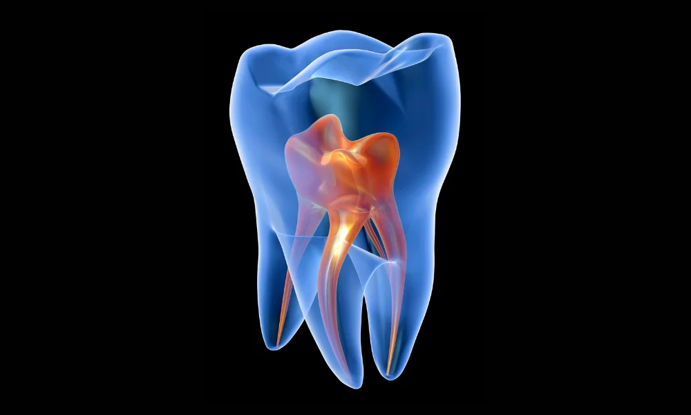
Root canal infections typically originate from bacterial infiltration into the pulp of the tooth. Common causes include:

  • Untreated Cavities: When cavities are left untreated, bacteria can penetrate the tooth, reaching the pulp.
  • Tooth Trauma: Physical injuries to the tooth can create openings for bacteria to enter and cause an infection.
  • Faulty Dental Procedures: Poorly performed dental procedures or inadequate fillings may leave room for bacterial entry.

Symptoms of Root Canal Infections

Recognizing the signs of a root canal infection is crucial for timely intervention. Symptoms may include:

  • Severe Toothache: Persistent and intense pain in the affected tooth is a common indicator.
  • Sensitivity to Hot or Cold: Discomfort when consuming hot or cold foods and beverages.
  • Swollen Gums: Inflammation and tenderness in the surrounding gums.
  • Pimple on the Gums: Formation of a pimple-like bump, known as a dental abscess, indicating infection.

Diagnosis and Treatment

Proper diagnosis involves dental examinations and imaging, often using X-rays. Once diagnosed, various treatment options are available:

  • Root Canal Therapy: The infected pulp is removed, and the canal is cleaned, disinfected, and sealed.
  • Apicoectomy: In cases of persistent infection, a surgical procedure to remove the apex of the tooth’s root may be necessary.
  • Extraction: In severe cases, if the tooth cannot be saved, extraction might be recommended.

Preventing Root Canal Infections

Maintaining good oral hygiene is paramount in preventing root canal infections:

  • Regular Dental Check-ups: Routine dental visits can help catch and address issues before they escalate.
  • Timely Treatment: Addressing cavities and dental trauma promptly reduces the risk of infection.
  • Proper Dental Care: Brushing, flossing, and using an antiseptic mouthwash contribute to overall oral health.

Understanding root canal infections empowers individuals to recognize symptoms early and seek timely dental care. With proper diagnosis and treatment, the discomfort associated with root canal infections can be alleviated, promoting oral health and overall well-being.

Also read: A Comprehensive Guide Through Root Canal X-rays

Types of Root Canal Infections and Their Causes

Root canal infections occur when bacteria invade the innermost part of the tooth, leading to inflammation and damage to the pulp tissue. These infections are often painful and, if left untreated, can result in abscesses, tooth loss, or more severe complications. Understanding the types of root canal infections and their causes is important in recognizing symptoms early and seeking appropriate treatment.

Types of Root Canal Infections

Root canal infections can be classified based on the extent of the damage to the pulp and surrounding tissues. The following are the common types:

1. Primary Endodontic Infection

A primary endodontic infection occurs when bacteria enter the root canal system for the first time, causing inflammation and infection. The infection is generally caused by deep dental decay, trauma, or cracks in the tooth that allow bacteria to penetrate the pulp.

  • Signs and Symptoms:
  • Severe toothache, especially when biting or chewing.
  • Sensitivity to hot or cold.
  • Swelling in the surrounding gum tissue.
  • Darkening of the tooth.

2. Secondary (Persistent) Endodontic Infection

A secondary or persistent endodontic infection arises after a previous root canal treatment. In this case, bacteria remain in the root canal system or reinvade the tooth, causing the infection to persist or reoccur. This type of infection is often due to incomplete cleaning of the canals during the initial treatment or reinfection due to a poor seal.

  • Signs and Symptoms:
  • Recurring pain in a previously treated tooth.
  • Swelling or tenderness in the gums near the tooth.
  • Discharge of pus or an abscess around the root.
  • Sensitivity to pressure or touch.

3. Acute Apical Periodontitis

Acute apical periodontitis is a root canal infection that spreads beyond the tooth and affects the periapical tissues (the area around the root tip). This infection often results in an inflammatory response in the surrounding bone, leading to increased pressure and pain.

  • Signs and Symptoms:
  • Intense, localized pain that is aggravated by pressure or chewing.
  • Tenderness in the gum or jawbone near the affected tooth.
  • Swelling and redness in the gum tissue.
  • In some cases, fever and general malaise.

4. Chronic Apical Periodontitis

Chronic apical periodontitis is a long-standing infection of the root canal system that often progresses without significant symptoms. The infection gradually destroys the surrounding bone and tissue, forming an abscess or cyst at the root tip.

  • Signs and Symptoms:
  • Mild or no pain, even though the infection is present.
  • Presence of an abscess or small bump (fistula) on the gum, which may drain pus.
  • Gradual bone loss visible on X-rays.
  • Occasional discomfort or swelling.

5. Acute Apical Abscess

An acute apical abscess is a severe and painful form of root canal infection characterized by the rapid accumulation of pus at the root tip. This infection typically occurs when the tooth’s pulp has become necrotic (dead) and bacteria spread into the surrounding tissues, causing inflammation and pus formation.

  • Signs and Symptoms:
  • Intense, throbbing pain that worsens quickly.
  • Swelling in the gum or face near the infected tooth.
  • Fever, feeling unwell, and difficulty opening the mouth (in severe cases).
  • A noticeable, pus-filled swelling on the gum or near the jaw.

6. Chronic Apical Abscess

A chronic apical abscess is a long-term infection that typically results from untreated decay or a failed root canal treatment. This condition may not cause significant pain, but it leads to the formation of a draining sinus or fistula. The abscess may release pus through the fistula, relieving pressure and reducing pain.

  • Signs and Symptoms:
  • Little to no pain, but pus drainage through a fistula.
  • Periodic swelling or discomfort in the affected area.
  • Bad taste in the mouth due to the discharge of pus.
  • Radiographic evidence of bone loss or cyst formation.

Causes of Root Canal Infections

Several factors can contribute to the development of root canal infections, with bacteria being the primary culprit. The following are the common causes:

1. Deep Tooth Decay

  • When decay penetrates through the enamel and dentin layers, bacteria can reach the pulp, leading to infection. Deep cavities allow bacteria to invade the root canal system, causing inflammation and infection of the pulp tissue.

2. Trauma or Injury

  • Cracked or fractured teeth caused by injury, excessive force, or grinding (bruxism) can expose the pulp to bacteria. Even if a tooth does not break entirely, trauma can damage the pulp and increase the risk of infection.

3. Failed or Incomplete Dental Work

  • A previously treated tooth may develop a secondary infection if the root canals were not fully cleaned or if the filling or crown does not seal the tooth properly. Bacteria can enter the tooth and cause reinfection, especially if cracks or leaks occur around a restoration.

4. Periodontal (Gum) Disease

  • Advanced gum disease can cause the gum tissue to recede, exposing the tooth roots to bacteria. In severe cases, bacteria can travel from the gums to the root canal system, causing a root canal infection.

5. Pulp Necrosis

  • When the pulp tissue inside the tooth dies (necrosis) due to decay or trauma, it creates an ideal environment for bacterial growth. This dead tissue can cause infection, which may spread to the surrounding bone and tissues.

6. Inadequate Oral Hygiene

  • Poor oral hygiene practices, such as infrequent brushing and flossing, allow plaque and bacteria to accumulate on the teeth and gums. Over time, this can lead to decay, infection, and root canal issues.

Proper oral hygiene, regular dental check-ups, and prompt treatment of decay or injuries are key to avoiding root canal infections. If symptoms arise, such as tooth pain or swelling, it’s essential to seek dental care to prevent the infection from worsening.

Anatomy of a Tooth and the Root Canal System: A Comprehensive Overview

Understanding the anatomy of a tooth and its root canal system is essential to appreciate how dental issues, such as decay or infection, develop and how treatments like root canals can resolve them. A tooth is much more than what we see on the surface; it has multiple layers and a complex internal structure that plays a critical role in its function and health.

The Anatomy of a Tooth

A tooth consists of two primary sections: the crown (the visible part above the gums) and the root (embedded in the jawbone, anchoring the tooth). Each section is made up of several distinct layers, each with its own function.

1. Enamel

  • Location: Outermost layer of the crown.
  • Composition: The hardest substance in the human body, made primarily of mineralized calcium phosphate crystals.
  • Function: Protects the inner layers of the tooth from decay and damage. While it is very strong, enamel can be eroded by acids in food and drinks, or damaged by trauma.

2. Dentin

  • Location: Beneath the enamel and cementum (in the root).
  • Composition: A less mineralized tissue than enamel, dentin has microscopic tubules that connect to the innermost part of the tooth.
  • Function: Supports the enamel and transmits sensations such as temperature changes or pressure. Dentin is more sensitive than enamel and is prone to decay if exposed.

3. Cementum

  • Location: Covers the root of the tooth, below the gum line.
  • Composition: A hard, calcified tissue that is less durable than enamel.
  • Function: Attaches the tooth to the periodontal ligament, helping to anchor the tooth to the bone. Cementum provides protection to the root and is essential for tooth stability.

4. Pulp

  • Location: The innermost part of the tooth, found in the pulp chamber and extending down through the root canals.
  • Composition: Soft tissue that contains nerves, blood vessels, and connective tissue.
  • Function: Supplies nutrients to the tooth and responds to stimuli such as pain, heat, and cold. The pulp is essential for the early development of the tooth but becomes less critical once the tooth has fully formed.

The Root Canal System

The root canal system lies beneath the surface of the tooth and is a key part of the tooth’s structure. This system includes the pulp chamber in the crown and the root canals that extend down into the roots, ultimately connecting to the jawbone.

1. Pulp Chamber

  • Location: The central cavity inside the crown of the tooth.
  • Function: Contains the tooth’s nerve and blood supply. The pulp chamber narrows into the root canals that extend toward the root tip.

2. Root Canals

  • Location: Narrow passageways that run from the pulp chamber down to the root tip, within each tooth root.
  • Function: The root canals house the tooth’s pulp (nerves, blood vessels), which keeps the tooth nourished and alive. The canals allow nutrients and sensory signals to travel to and from the tooth.

Teeth may have one or more root canals, depending on their type:

  • Incisors and canines typically have one root canal.
  • Premolars may have one or two root canals.
  • Molars often have two to four root canals, with complex branching in their root structures.

Importance of the Root Canal System in Dental Health

The root canal system is crucial for the tooth’s vitality, but it is also where problems like infections can develop. When bacteria invade the tooth, typically through deep decay or cracks, the pulp becomes infected or inflamed. This infection can spread through the root canal system, causing pain, swelling, and eventually leading to an abscess if left untreated.

Common Issues Affecting the Root Canal System

  • Pulpitis: Inflammation of the pulp caused by decay, trauma, or bacterial infection. Pulpitis can cause sharp, localized tooth pain.
  • Abscess: A severe infection that occurs when bacteria reach the root tip and cause pus to collect in the surrounding bone.
  • Necrotic Pulp: If the infection is not treated, the pulp can die (necrosis), leading to the need for a root canal procedure or extraction.

Root Canal Treatment

If the root canal system becomes infected or damaged, a root canal procedure may be necessary. This treatment involves:

  1. Removing the infected pulp: The dentist or endodontist removes the damaged or infected pulp from the tooth.
  2. Cleaning the canals: The empty canals are thoroughly cleaned to eliminate any remaining bacteria or debris.
  3. Filling the canals: The cleaned root canals are filled with a rubber-like material called gutta-percha to seal them off and prevent reinfection.
  4. Sealing the tooth: A filling or crown is placed on the tooth to restore its structure and function.

Root canal treatment preserves the tooth by removing the damaged tissue while maintaining the tooth’s structural integrity. Without this treatment, the infection could spread, leading to tooth loss or other complications.

When problems arise in the root canal system, timely treatment such as a root canal can save the tooth and prevent further complications. Understanding these structures helps patients better appreciate their dental health and the importance of preventive care.

Why X-rays Are Important for Diagnosing Root Canal Infections

X-rays are a critical tool in diagnosing root canal infections. While a dentist can examine your tooth and identify signs of infection, an X-ray allows them to see below the surface and assess the condition of the tooth’s roots and surrounding bone.

How X-rays Help in Diagnosis

  • Detect hidden infections: In some cases, the infection may not be visible to the naked eye. X-rays can reveal areas of decay or infection that are otherwise hidden.
  • Examine the root structure: Root canal infections often affect the tooth’s root, which is difficult to assess without an X-ray. The X-ray provides a detailed view of the root canals and the extent of the infection.
  • Assess bone health: A root canal infection can cause bone loss around the affected tooth. X-rays help the dentist determine if the bone has been damaged and how extensive the damage is.
  • Guide treatment decisions: The X-ray gives the dentist important information about the severity of the infection, helping them decide whether a root canal treatment or extraction is necessary.

Types of X-rays Used for Root Canal Diagnosis

There are several types of dental X-rays, each serving a different purpose. For root canal diagnosis, the most common types of X-rays are:

1. Periapical X-rays

A periapical X-ray provides a detailed image of the entire tooth, including the crown, roots, and surrounding bone. This type of X-ray is typically used to assess the health of the tooth’s roots and check for signs of infection, abscesses, or bone loss.

When Is It Used?

  • To examine the tooth’s root structure
  • To detect infections or abscesses at the tip of the root
  • To evaluate bone health around the tooth

2. Panoramic X-rays

Panoramic X-rays capture the entire mouth in one image, including all teeth, the upper and lower jaws, and the sinuses. While less detailed than periapical X-rays, panoramic X-rays provide a broad view of the oral cavity and can help detect larger infections or issues affecting multiple teeth.

When Is It Used?

  • To get a comprehensive view of the entire mouth
  • To assess the jawbone and surrounding structures
  • To detect widespread infections or abnormalities

3. Cone Beam Computed Tomography (CBCT)

Cone beam computed tomography (CBCT) is a more advanced type of X-ray that provides a 3D image of the tooth and surrounding structures. CBCT scans are particularly useful for complex cases where a detailed view of the tooth’s anatomy is required. This type of X-ray is often used before performing a root canal procedure to guide the dentist during treatment.

When Is It Used?

  • For detailed examination of the tooth’s internal structure
  • To plan complex root canal treatments
  • To assess the extent of the infection in 3D

The X-ray Process

If your dentist suspects a root canal infection, they will likely take an X-ray to confirm the diagnosis. Here’s what you can expect during the X-ray process:

Step 1: Preparation

Before the X-ray, you’ll be asked to remove any jewelry or metal objects from your head and neck area. A lead apron will be placed over your chest and abdomen to protect you from unnecessary radiation exposure.

Step 2: Taking the X-ray

The dentist or dental assistant will position the X-ray machine near your mouth. You’ll either bite down on a small piece of plastic to hold the film in place or stand still in front of the machine for a panoramic X-ray. The X-ray itself only takes a few seconds, and you won’t feel any discomfort during the process.

Step 3: Reviewing the Results

After the X-ray is taken, the dentist will review the images and discuss the findings with you. If an infection is present, they’ll explain the extent of the damage and recommend the best course of treatment, whether that’s a root canal procedure or another option.

Risks and Safety of Dental X-rays

Dental X-rays are generally considered safe, as they use very low levels of radiation. However, some patients may be concerned about the potential risks of radiation exposure. Here’s what you need to know:

Low Radiation Levels

The amount of radiation used in dental X-rays is minimal, especially with modern digital X-ray machines. The risk of harm is extremely low, but your dentist will still take precautions, such as using a lead apron, to minimize exposure.

Frequency of X-rays

The frequency of dental X-rays will depend on your specific dental needs. If you’re experiencing symptoms of a root canal infection, an X-ray is essential for an accurate diagnosis. Your dentist will only recommend X-rays when necessary to ensure your safety.

Special Considerations for Pregnant Patients

If you’re pregnant or think you might be, it’s important to let your dentist know before getting an X-ray. In most cases, dental X-rays are safe during pregnancy, but your dentist may choose to delay non-urgent X-rays until after your baby is born.

Treatment Options After X-ray Diagnosis

Once your dentist confirms a root canal infection through X-rays, they’ll recommend the appropriate treatment based on the severity of the infection. Common treatment options include:

Root Canal Treatment

A root canal treatment is a procedure that removes the infected pulp from the tooth, cleans the root canals, and seals them to prevent further infection. This treatment is highly effective in saving the affected tooth and relieving pain.

Tooth Extraction

In cases where the infection is too severe or the tooth is badly damaged, extraction may be necessary. Your dentist will remove the infected tooth to prevent the infection from spreading to surrounding teeth and bone.

Antibiotics

In some cases, antibiotics may be prescribed to help control the infection, especially if there is swelling or if the infection has spread beyond the tooth. Antibiotics are typically used in conjunction with root canal treatment or extraction.

Benefits of X-ray Diagnosis

Accuracy in Identification

X-rays enable dentists to identify root canal infections accurately, distinguishing them from other dental issues with similar symptoms.

Treatment Customization

Precise diagnosis allows for tailored treatment plans, addressing the specific nature and extent of the root canal infection.

Monitoring Progress

X-rays aid in monitoring the progress of treatments, ensuring the elimination of infection and the success of interventions.

Going Beyond Symptoms

Symptom Ambiguity

Root canal infections can manifest as various symptoms, making X-rays crucial for accurate diagnosis in cases where symptoms alone may be ambiguous.

Hidden Infections

Some infections may not exhibit noticeable symptoms, emphasizing the importance of X-rays in detecting hidden or asymptomatic infections.

Unveiling Hidden Realities

When dealing with a root canal infection, a profound understanding goes beyond just addressing surface-level symptoms. The use of X-ray imaging emerges as a pivotal tool, allowing dental professionals to delve into concealed complexities and make informed decisions for comprehensive oral health.

Beyond the Surface

Root canal issues often hide beneath the visible symptoms, requiring a more in-depth exploration. X-ray images play a crucial role in providing a detailed analysis of tooth structures. They unveil hidden infections, showcase intricate canal anatomy, and highlight potential challenges that might not be immediately apparent during a routine examination.

How to Diagnose Patient from X-ray of Root Canal Infection?
Revealing the Unseen

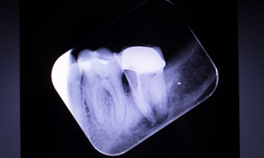
Diagnosing a root canal infection requires a meticulous approach, and X-ray analysis stands as a crucial diagnostic ally. Beyond surface symptoms, these images unveil the unseen, providing an intricate view of the tooth’s interior.

Identifying Infections and Anatomy

X-rays highlight the presence and extent of infections, showcasing the affected areas within the root canal system. Moreover, they illuminate the intricate anatomy of the canals, helping dentists navigate complexities that may impact the treatment approach.

Precision in Treatment Planning

Armed with the insights gleaned from X-ray analysis, dentists can precisely plan the treatment. Whether it’s determining the extent of infection, locating additional canals, or assessing potential complications, this in-depth diagnosis sets the stage for a tailored and effective treatment strategy.

Enhancing Patient Care

The ability to diagnose root canal infections through X-ray analysis goes beyond detection; it enhances overall patient care. It allows for early intervention, reduces the risk of complications, and ensures that treatment is targeted, leading to more successful outcomes in managing and treating root canal infections.

Practical Tips to Prevent Root Canal Infections and Maintain Good Oral Health

Preventing root canal infections and maintaining optimal oral health are essential for preserving your teeth and avoiding painful and costly dental procedures. By following a consistent oral hygiene routine and making smart lifestyle choices, you can reduce the risk of tooth decay, gum disease, and root canal infections. Here are some practical tips to keep your teeth healthy and minimize the chances of developing a root canal infection.

1. Brush Twice a Day with Proper Technique

Brushing your teeth twice a day is the foundation of good oral hygiene. Proper brushing helps remove plaque, bacteria, and food particles that can lead to tooth decay and gum disease.

  • Tip: Use a soft-bristled toothbrush and fluoride toothpaste. Hold the brush at a 45-degree angle to your gums, and gently brush all surfaces of your teeth (front, back, and chewing surfaces) in small, circular motions.
  • Timing: Spend at least two minutes brushing to ensure that you clean all areas thoroughly.

2. Floss Daily to Remove Plaque Between Teeth

Flossing is just as important as brushing, as it removes plaque and food particles from areas that your toothbrush can’t reach—between the teeth and under the gumline. Plaque buildup in these areas can lead to cavities and gum disease, increasing the risk of root canal infections.

  • Tip: Gently slide the floss between your teeth, following the curve of each tooth. Be careful not to snap the floss into the gums, as this can cause irritation.

3. Use an Antibacterial Mouthwash

Mouthwash can help reduce the bacteria that cause plaque, tooth decay, and gum disease, giving your mouth an extra layer of protection. An antibacterial mouthwash also freshens your breath and reaches areas that brushing and flossing might miss.

  • Tip: Choose a mouthwash with fluoride or antibacterial ingredients to strengthen your enamel and reduce plaque buildup. Rinse for 30 seconds after brushing and flossing.

4. Maintain a Healthy Diet

What you eat plays a major role in your oral health. A diet high in sugar and acidic foods can contribute to tooth decay, which is a common cause of root canal infections. To keep your teeth strong and healthy, focus on nutrient-rich foods that support your overall well-being.

  • Tip: Limit sugary snacks, sodas, and acidic drinks like citrus juices, which can erode enamel and lead to cavities. Instead, eat a balanced diet that includes plenty of fruits, vegetables, lean proteins, and dairy products.
  • Calcium: Foods rich in calcium, such as milk, yogurt, and cheese, help strengthen your teeth and prevent decay.

5. Stay Hydrated with Water

Water not only keeps your body hydrated but also promotes saliva production, which is important for maintaining a healthy mouth. Saliva neutralizes acids, washes away food particles, and helps prevent the buildup of plaque on your teeth.

  • Tip: Drink water throughout the day, especially after meals. Avoid sugary beverages like sodas and sports drinks, as they can promote tooth decay and increase the risk of root canal infections.

6. Don’t Ignore Early Signs of Dental Problems

Tooth pain, sensitivity to hot or cold, and swelling in the gums are early warning signs of potential dental issues. Ignoring these symptoms could allow minor problems to escalate into serious infections requiring root canal treatment.

  • Tip: If you experience tooth pain or notice any unusual symptoms, visit your dentist as soon as possible. Early treatment can prevent the need for more invasive procedures.

7. Visit Your Dentist Regularly

Routine dental check-ups are essential for maintaining good oral health. Your dentist can identify early signs of tooth decay, gum disease, and other issues before they progress to more serious conditions like root canal infections.

  • Tip: Schedule a dental check-up every six months. Your dentist will perform a thorough cleaning to remove plaque and tartar, and they will examine your teeth for any signs of decay, cracks, or infection.

8. Consider Dental Sealants

Dental sealants are a protective coating applied to the chewing surfaces of your back teeth (molars), where decay often starts. This preventive measure can help reduce the risk of cavities, especially in children and adults with deep grooves in their teeth.

  • Tip: Talk to your dentist about whether dental sealants are right for you or your children. Sealants are a simple and effective way to prevent decay in vulnerable areas of the mouth.

9. Quit Smoking and Avoid Tobacco Products

Smoking and using tobacco products significantly increase the risk of gum disease and tooth decay, both of which can lead to root canal infections. Tobacco use also weakens your immune system, making it harder for your body to fight infections and heal after dental treatments.

  • Tip: If you smoke or use tobacco, consider quitting to improve your oral and overall health. There are many resources available to help you quit, including support groups and nicotine replacement therapies.

10. Manage Bruxism (Teeth Grinding)

Bruxism, or teeth grinding, can wear down the enamel and lead to cracks, chips, and fractures. These issues can expose the inner layers of the tooth to bacteria, increasing the risk of a root canal infection.

  • Tip: If you grind your teeth, especially at night, talk to your dentist about getting a custom mouthguard. This will help protect your teeth from damage and prevent future problems.

By practicing consistent oral hygiene, eating a balanced diet, and visiting your dentist regularly, you can protect your teeth and avoid painful infections. If you experience any signs of dental issues, don’t hesitate to seek professional care—early intervention is the key to long-term dental health.

The Importance of Early Diagnosis and Treatment of Root Canal Infections to Prevent Complications

Root canal infections are not only painful but can lead to serious complications if left untreated. Early diagnosis and timely treatment are crucial to prevent the spread of infection and preserve the health of your tooth. In this section, we will explore why early detection is vital and how prompt treatment can save your tooth and prevent further dental issues.

1. Prevents the Spread of Infection

A root canal infection typically begins when bacteria enter the pulp chamber through a cavity, crack, or trauma to the tooth. If not treated in time, the infection can spread beyond the tooth’s root to the surrounding tissues, gums, and even the jawbone.

  • Complications: Once the infection spreads, it can result in abscesses, which are pockets of pus that form at the base of the tooth. These abscesses are painful and can cause swelling, fever, and difficulty chewing. If untreated, the infection can spread to other parts of the body, including the bloodstream, leading to more severe health risks like sepsis.

2. Saves the Tooth

When root canal infections are detected early, the tooth can often be saved through a root canal procedure, which removes the infected pulp and seals the tooth to prevent reinfection. If the infection is allowed to progress, the damage to the tooth may become so severe that extraction becomes the only option.

  • Benefit: Saving your natural tooth is always preferable to extraction. Your natural teeth maintain the integrity of your jawbone and bite alignment, and they are easier to maintain than artificial replacements.

3. Avoids Tooth Loss and Costly Dental Procedures

Treating a root canal infection early is far less invasive and costly than dealing with complications from an untreated infection. If the infection causes significant damage, you may require a tooth extraction, followed by a dental implant, bridge, or denture to restore functionality. These procedures are more expensive and time-consuming than a root canal treatment.

  • Benefit: Early intervention helps you avoid the expense and discomfort of more complex dental procedures that become necessary when infections are not addressed promptly.

4. Prevents Bone Loss

If an infection spreads to the jawbone, it can cause bone deterioration. This can weaken the structure of your jaw and lead to more extensive dental and medical problems. Bone loss can affect your facial structure and cause further complications with neighboring teeth.

  • Impact: Treating the infection before it reaches the bone helps preserve the health of your jawbone and supports your overall oral health. Once bone loss occurs, it may require additional treatments like bone grafts to rebuild the affected areas.

5. Prevents Gum Disease

An untreated root canal infection can also contribute to gum disease. As the infection spreads to the gums, it causes inflammation and damage to the surrounding tissues. Gum disease can result in receding gums, loose teeth, and further bone damage.

  • Tip: Addressing root canal infections early not only saves the tooth but also prevents the onset or worsening of gum disease, which is another major cause of tooth loss.

6. Relieves Pain and Discomfort

One of the earliest signs of a root canal infection is pain, which can range from mild sensitivity to severe throbbing. The longer you wait to seek treatment, the more intense and frequent the pain will become. Early diagnosis allows for prompt treatment, alleviating discomfort and restoring your quality of life.

  • Benefit: Timely treatment of root canal infections not only saves your tooth but also eliminates the pain and discomfort associated with the infection.

7. Restores Proper Functionality

A root canal infection can make it difficult to chew, eat, and even speak properly. Once the infection is treated and the tooth is restored with a filling or crown, you can resume normal oral functions without pain or difficulty.

  • Tip: Early treatment ensures that the tooth remains functional and avoids the need for more complex restoration methods that may be required for severely damaged teeth.

Addressing the infection as soon as possible can save your tooth, protect surrounding tissues, and improve your overall oral health. Regular dental check-ups, along with swift action at the first sign of pain or discomfort, are key to catching and treating root canal infections early.

Conclusion

X-rays play a crucial role in diagnosing root canal infections, providing dentists with detailed images of the tooth’s internal structure and surrounding bone. With the help of X-rays, dentists can accurately assess the extent of the infection and recommend the most effective treatment options.

If you’re experiencing symptoms of a root canal infection, don’t wait to seek treatment. Early diagnosis and intervention can save your tooth and prevent the infection from spreading. Talk to your dentist about getting an X-ray to ensure a precise diagnosis and appropriate care.

References

Share:

Facebook
Twitter
Pinterest
LinkedIn